Resources
 Part of the Oxford Instruments Group
Expand
Collapse
Part of the Oxford Instruments Group
Application Notes
Author: Dr. Björn Braunschweig, Friedrich-Alexander University Erlangen-Nürnberg Institute of Particle Technology (LFG)
Published: 01 Jul 2015 · Last updated: 16 Jul 2025
Tags: nl-spectroscopy-app
In situ investigations of surfaces and interfaces with optical spectroscopy are often highly challenging because many linear optical methods do not have the ability to separate the signal generated at the interface from the signal of the bulk material. In fact, in these cases the signal from the interface is being overwhelmed by the bulk signal that is because there are several orders of magnitude more atoms/molecules in the bulk compared to the molecules/atoms at the interface. Obviously, in interface spectroscopy the experimenter faces two challenges which are selectivity and sensitivity.
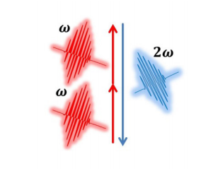
Figure 1 Schematic diagram of the three-photon process in SHG.
However, second-order nonlinear optical techniques such as second-harmonic (SHG) [1-3] and sum-frequency generation (SFG) [4-5] have been shown to be the ideal tools to face these challenges. That is because the three-photon processes of SFG and SHG are for materials with inversion symmetry such as liquids, gases, cubic crystals etc. forbidden in the bulk and only allowed at the surface or interfaces of these materials which necessarily represent breaks in the prevailing bulk symmetry. For that reason SFG and SHG can be used as experimental tools to study the structure of interfaces on a molecular level.
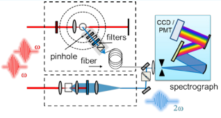
Figure 2 Schematic diagram of a setup for second-harmonic generation at colloidal interfaces. Not shown is the Ti:Sapphire based laser system for generation of tunable femtosecond laser pulses.
Since the conversion of the fundamental wave into the second harmonic is extraordinary small (≈10-11) due to the few contributing molecular layers, a highly efficient detection system is required besides intense laser excitation with femtosecond light pulses. For the detection of SHG one typically applies either – low-noise CCD cameras or photo multipliers (Figure 2). In order to spectrally resolve SHG photons and photons from possible multi-photon fluorescence processes, a combination of a spectrograph with one or two CCD cameras is most suited. In our current setup we use a combination of a Shamrock 303i 300 mm spectrograph (Andor Shamrock 303i) that is equipped with two CCD detectors (Andor Newton DU920P-BU2 and DU920P-BU) at its exit ports. The use of two CCDs enables us to optimize the quantum efficiency between 200 and 300 nm with the DU920P-BU2 model and between 300 and 700 nm with the DU920P-BU model.
We can collect SHG signals either by hard coupling with reflective optics or by fiber optics into the detection systems. The latter allows us to switch easily between different experimental setups e.g. SHG at planar interfaces or SHG at colloidal interfaces where we also measure the SHG intensity as a function of the scattering angle. For the latter we use a multi-leg fiber bundle which allows us to pick up SHG signals at different positions around our sample (Figures 2) and/or from a reference sample. With the spectrograph/CCD combination we are able to measure the generated signals spectrally resolved at different tracks on the CCD chip without significant cross talk (Figure 3a). Figure 3b shows photographs of our experimental setup that includes the laser system for SHG excitation and the detection system as described above.
Figure 4 demonstrates the importance to resolve the SHG intensity spectrally for colloidal Au nanoparticles which were stabilized by a citrate surface layer. The narrow feature at 400 nm originates from the second harmonic wave of the fundamental beam which was centered at a wavelength of 800 nm. The broad contribution is due to two- photon fluorescence presumably from an Au electronic state into the LUMO of the citrate adsorbate. Similar effects can be seen for colloidal quantum dots such as ZnS where two-photon fluorescence is also observed.
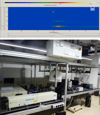
Figure 3 (a) Image of the CCD and spectra from three different sources (SHG reference and two photon luminescence from different sample positions). (b) Photograph of the femtosecond laser system for resonant SHG and the detector consisting of spectrograph and CCD cameras (right hand side).
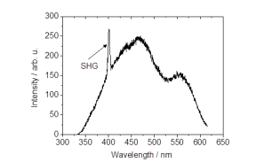
Figure 4 Spectrally resolved SHG signal from colloidal Au nanoparticles with a nominal size of 15 nm. For the excitation, laser pulses of 80 fs duration, a wavelength of 800 nm and a bandwidth of 10 nm FWHM were used. The intensity was detected under a scattering angle of 90°.
[1] C. Sauerbeck, M. Haderlein, B. Schürer, B. Braunschweig, W. Peukert and R.N. Klupp Taylor; Shedding Light on the Growth of Au Nanoshells; ACS Nano 8, 3088-3096 (2014).
[2] C. Sauerbeck, B. Braunschweig and W. Peukert, Surface charging and interfacial water structure of amphoteric colloidal particles, J. Phys. Chem. C 118, 10033-10042 (2014).
[3] S. Bergfeld, B. Braunschweig and W. Daum. Nonlinear optical spectroscopy of suboxides at oxidized Si(111) interfaces; Phys. Rev. Lett.,93, 097402-1-4 (2004).
[4] K. Engelhardt, W. Peukert and B. Braunschweig; Vibrational sum-frequency generation at protein modified air-water interfaces: Effects of molecular structure and surface charging; Curr. Opinion Coll. Int. Sci.;19, 207-215 (2014).
[5] K. Engelhardt, U. Weichsel. E. Kraft, D. Segets, W. Peukert, and B. Braunschweig, Mixed Layers of β-Lactoglobulin and SDS at Air-Water Interfaces with Tunable Intermolecular Interactions; J. Phys. Chem. B; 118, 4098-4105 (2014).

© Oxford Instruments 2026
