

Compact - fits on a bench, no need for a dark room, built-in antivibration mechanism.
Resources
 Part of the Oxford Instruments Group
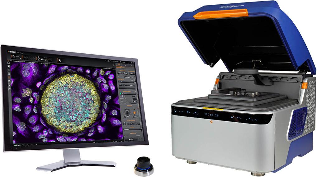
BC43 benchtop microscopes offer high-quality images, 2D/3D, widefield or confocal. The super-resolution option delivers resolutions down to 140 nm with the push of a button. Its certified quality ensures reproducibility and reliability of data between experiments and different research groups.
BC43 includes Fusion Benchtop software, for straightforward multimodal imaging acquisition, and Imaris, the market leader in image analysis software.
New: Discover BC43 microscopy for Bio-safety Labs.
Small in size, Big in performance.
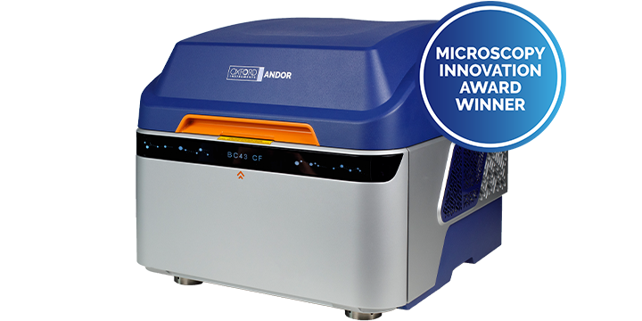
With certified quality, BC43 offers productivity and image excellence while maintaining the ease of use of a conventional entry-level widefield system, delivering high-end features at a competitive price.
Click the hotspots to see BC43 features and benefits explained.
Compact - fits on a bench, no need for a dark room, built-in antivibration mechanism.
Super-fast learning curve - new users can be independent within 30 min to 2 h – minimal training and user support needed.
Modular and upgradable - Widefield or Confocal, in-field upgrade , Modular software to adapt to user needs.
Unique & certified quality - IQ/OQ quality control - Certified by experts in the scientific imaging field QUAREP-LiMi*.
Target productivity - dual microlens spinning disk integrated with a sensitive sCMOS camera and specialised SW modules.
Live cell imaging - dual microlens spinning disk, efficient light collection results in gentle imaging, (image model organisms for days).
Versatility - with 4 lasers, 4 imaging modalities and a range of objectives, all your important microscopy lab experiments can be done hassle-free.
Image Analysis – Includes Imaris the gold standard software that delivers image analysis with precision, speed and user-friendly workflows.
* Consortium for Quality Assessment and Reproducibility for Instruments and Images in Light Microscopy
IQ/OQ is a unique and innovative service offering from Oxford Instruments that ensures the performance of all our benchtop microscope systems. Guaranteed, measured performance enables the comparison of experimental results across time and laboratories conducting the same research. Trust your data, validate your results.
“IQ OQ initiative holds great significance for scientific experiments in our field. It promotes standardised procedures, reduces variability, and enhances the reproducibility of results. Ultimately, it contributes to the credibility of research done with microscopes and advances the pursuit of scientific knowledge “
Dr. Glyn Nelson, Core Facility manager – Newcastle University, UK, QUAREP-LiMi Member
Learn More Request Pricing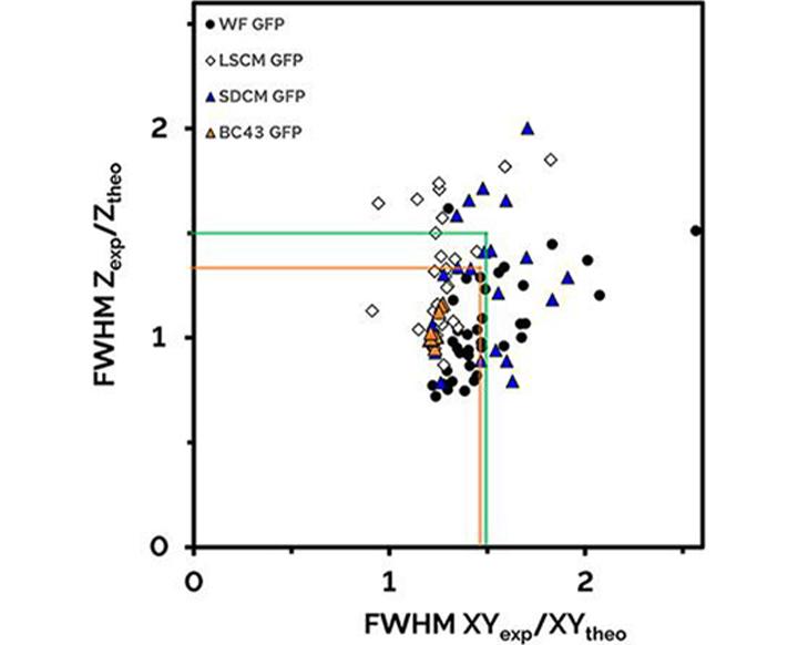
Lateral and axial resolutions of different types of fluorescent microscopes as a ratio of the theoretical full width half-maximum (FWHM).
Acceptable normalized resolution, within green square. Orange square shows that all measured BC43 (BC43 GFP) results are significantly better that the proposed spec of QUAREP-LIMi (green square). Note the variation observed on competing widefield (WF-GFP), laser scanning ( LSCM GFP) and spinning disk (SDCM GFP) competitors products. Figure adapted from data and specifications included in Faklariset al.
At the heart of BC43 is a software that is easy to use yet feature rich, flexible and modular, allowing the system to grow with your research needs. Fusion Benchtop includes features that enhance productivity and provide high-quality image visualisation.
Some of the fusion bench top features include:
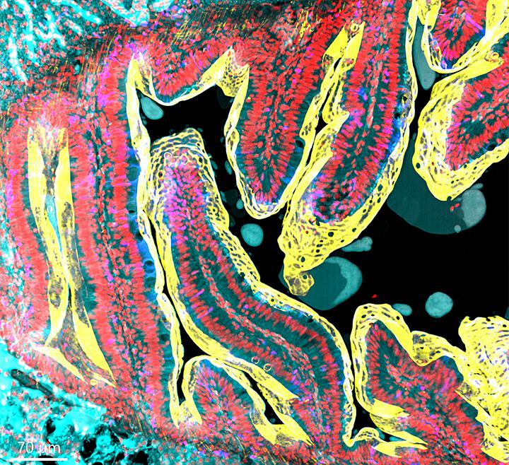
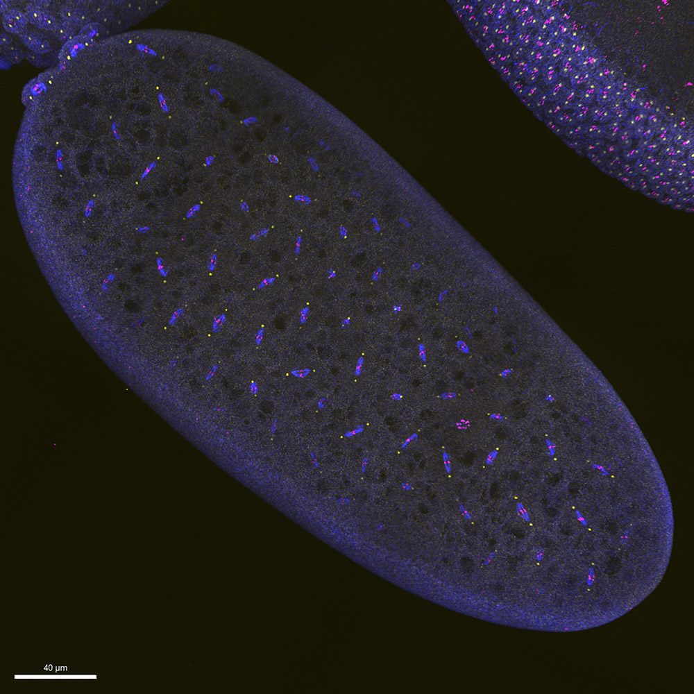
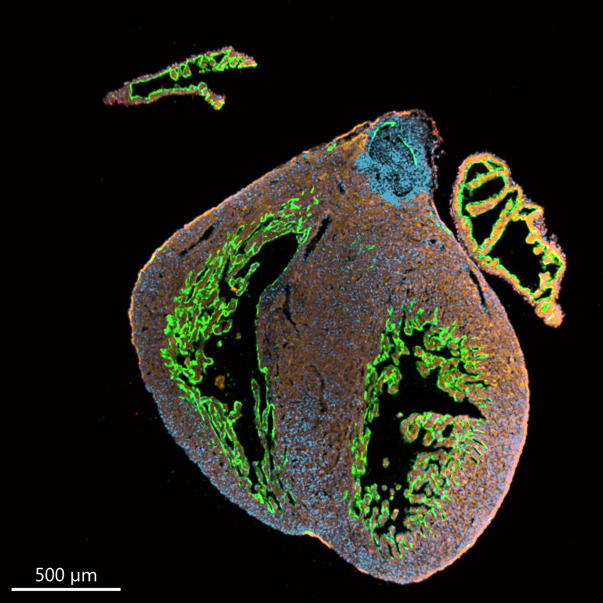
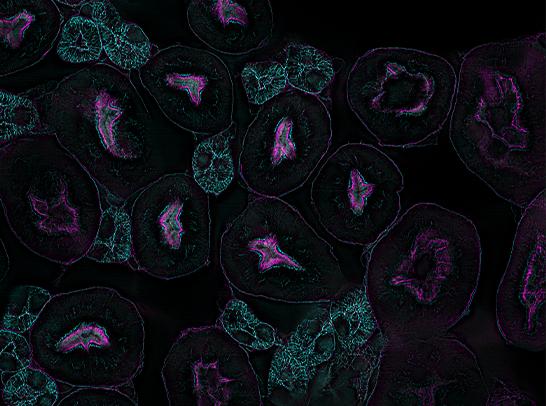
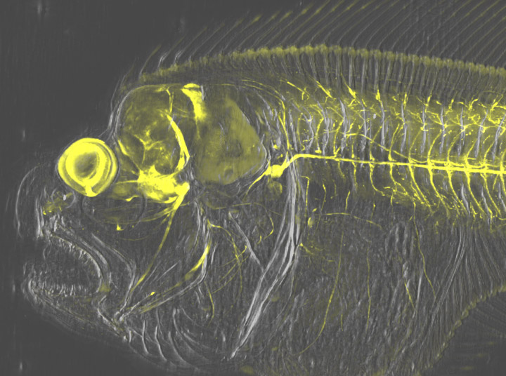
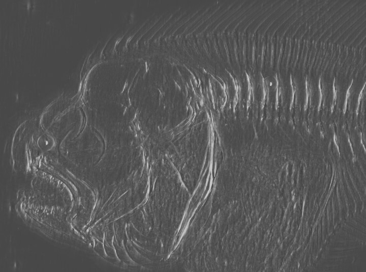
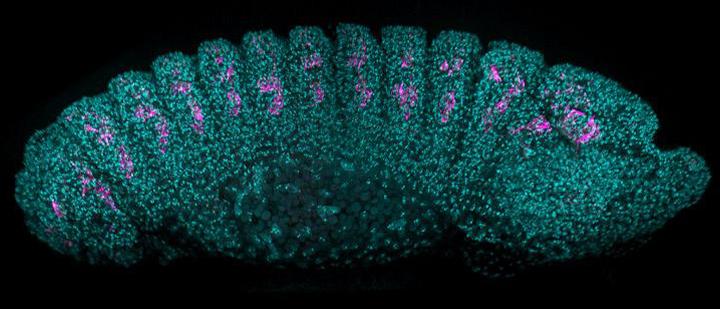
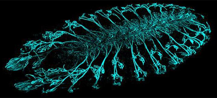
Zebrafish intestine stitched image.
Image was acquired using the confocal imaging modality of BC43, with 4 imaging channels, 77 stacks and 28 tiles. The full stitched image is composed of a total of 8624 images. The deconvolution and stitching options were both activated on the protocol. (image acquired ~ 30 min).
Sample courtesy of Julien Resseguier, at NorMic, University of Oslo. Image credit: Claudia Florindo, Oxford Instruments
All Andor Benchtop Microscopes come with the Imaris Quant package for users to:
Additional Imaris modules are also available including tools for tracking and reporting motility metrics, quantifying cells and their intracellular organelles, and tracing neurons for reporting their specific characteristics.
We image high numbers of thick and large tissue samples demanding a reliable confocal microscope to handle big experiments. BC43 is a true workhorse, requiring minimal experience to obtain high quality images. Being able to put it on a regular bench in our own lab is an efficiency bonus!
Professor Jacco van Rheenen, The Netherlands Cancer Institute.


Our research is focused on the cellular and molecular interactions taking place during normal heart development. To gain insight into these we rely heavily on imaging approaches and the BC43 confocal system has been instrumental to advance our studies. Its low footprint combined with its multi-imaging modalities and plug-and-play features are a perfect match!
Joaquim Miguel Nunes Vieira, PhD, King's College London.


© Oxford Instruments 2026
 Powered by Bioz