Resources
 Part of the Oxford Instruments Group
Expand
Collapse
Part of the Oxford Instruments Group
Application Notes
Author: Dr. Andrew P. Carpenter
Published: 01 May 2025 · Last updated: 29 Jul 2025
X-ray imaging and spectroscopy continues to yield innumerable contributions in material science, life science, geological science, and beyond. Looking towards the future – high energy sources (ex. Synchrotrons, XFELS, etc) possess a higher brilliance, higher repetition rates, and higher time resolution. These advances are paving the way for the development of new experimental methodologies and opening the door for the acquisition of large amounts of data. Concurrent with the development of these new high energy sources is the need for advances in X-ray camera technologies that can facilitate the high-throughput generation of data and be incorporated in complex experimental geometries. Importantly, it is critical these camera technologies do not compromise on sensitivity and accuracy of detection while exploiting the higher frame rates of these next generation sources to benefit time consuming applications (e.x. tomography).
We present several detection solutions for the new high energy sources of the future that are brighter and faster than previous generations. These detection solutions, based on sCMOS camera technology, include direct detection methods for the EUV-soft X-ray regimes and indirect detection methods extending sensitivity into the hard X-ray energies. Looking towards higher brilliance sources and high-throughput methodologies we highlight full frame acquisition rates an order of magnitude, or more, faster than comparable CCD technology while maintaining a high QE and lower noise floor. Further discussion around these cameras is framed in the context of how they can contribute to current experimental methodologies, quickly generate large amounts of data, and how they can facilitate the continual evolution in X-ray methodologies into the future.
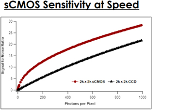
Signal-to-noise comparison of similarly
sized sCMOS (red) and CCD (black)
cameras during fast acquisition
operating conditions (100 fps).
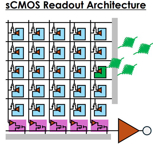
The change in readout architecture enables at least an order of magnitude higher frame rates!
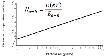
Multiple photoelectron generation in silicon detectors upon absorption of HE photons.
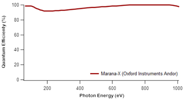
Quantum efficiency (%) of an HED, direct detection, sCMOS camera over the EUV and soft X-ray energy range.
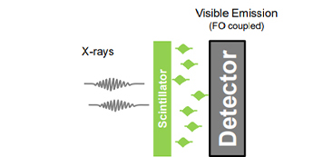
Indirect detection with scintillators coupled to sCMOS cameras, via fiber optic plates or lenses provide flexibility in scintillator choice, experimental design, and energy sensitivity. Both X-ray and neutron scintillators add to the diversity of application.
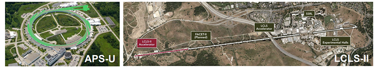
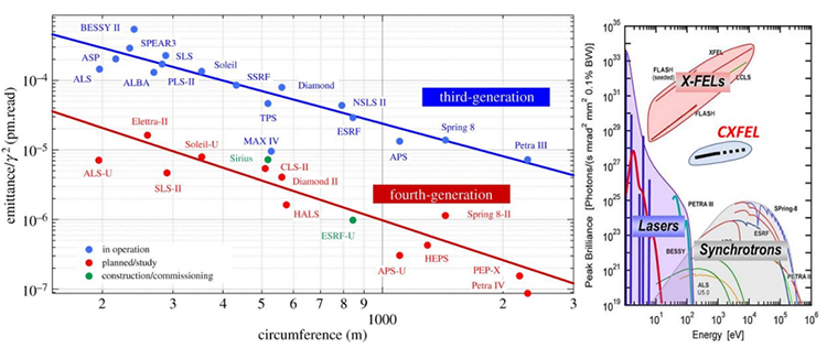
The new and upgraded synchrotrons (e.x. APS-U) are coming online with higher coherence (left) 1 and brilliance (right) 2 . Along with HHG lasers and X-ray free electron lasers (XFELS), high energy sources are getting brighter, with higher coherence, and faster rep rates.
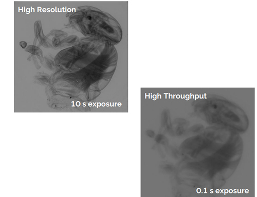
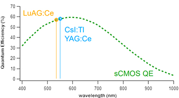
Variation of scintillator enables higher resolution versus higher throughput application. Both images recorded with a 40 keV X-ray source. Left image recorded using 20 µm YAG:Ce scintillator. Right image was acquired using a 150 µm CsI:Tl scintillator with a 0.1 second exposure. Images courtesy of Crytur.
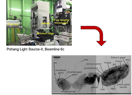
Negative phase contrast image of an ant recorded using 25 keV X-ray energy and a 100 ms exposure time.3
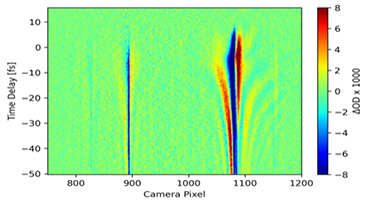
Non-colinear excitation of Argon using attosecond XUV pulse with two NIR pulses. Primary absorption line is observed for argon autoionization (25-30 eV) with multi-wave mixing features observed above and below.
The readout architecture of the direct detection sCMOS (Marana-X) avoids artifacts common in CCD detection (blooming and smearing), enabling more robust multi-wave mixing experiments.
Transient absorption spectrum characterizing Neon autoionization (~45 eV). Neon was excited by an attosecond XUV pulse that was colinear with a NIR pulse. Such a measurement can be useful for characterizing the crosscorrelation time between pulses and monitor an experimental system’s condition. Entire measurement took ~30 seconds as the exposure for each delay time lasted ~40 ms.
*Data courtesy of Dr. Lauren Drescher, recorded during Marana-X demo.
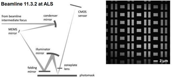
General description of the zoneplate based EUV microscope at ALS beamline 11.3.2 (left) and an example image recorded through photomask pellicle (right).4
EUV sensitivity, small pixel size, fast image readout, and sCMOS readout architecture enable experimental flexibility in imaging speed while maintaining high image quality, high contrast, and high signal to noise.
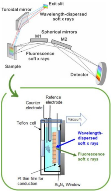
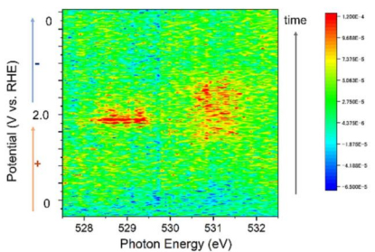
Real-time wavelength-dispersive XAS (left) was performed, at beamline BL16A of the Photon Factory in Japan, for real-time monitoring of oxygen species during the OER on a cobalt oxide electrocatalyst.
The oxygen K-edge spectra (below) were recorded as a function of electrochemical potential. Direct detection sCMOS readout speed and sensitivity enabled fast acquisition of these spectra.5
2. https://www.xtal.iqf.csic.es/Cristalografia/parte_13-en.html

© Oxford Instruments 2026
