Part of the Oxford Instruments Group
Expand

Image Acquisition and Analysis Solutions for Neuroscience

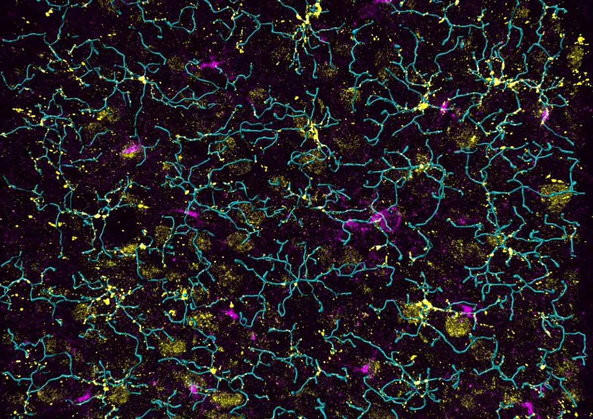
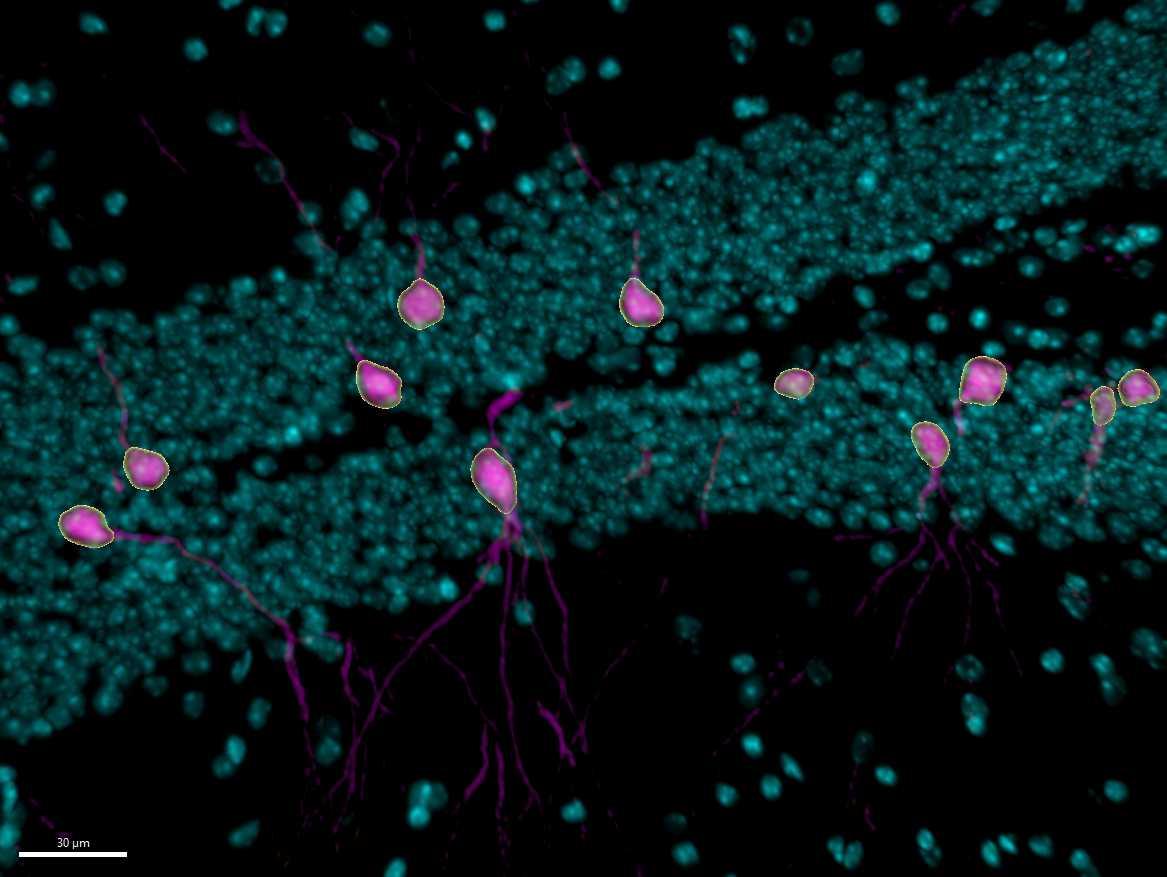
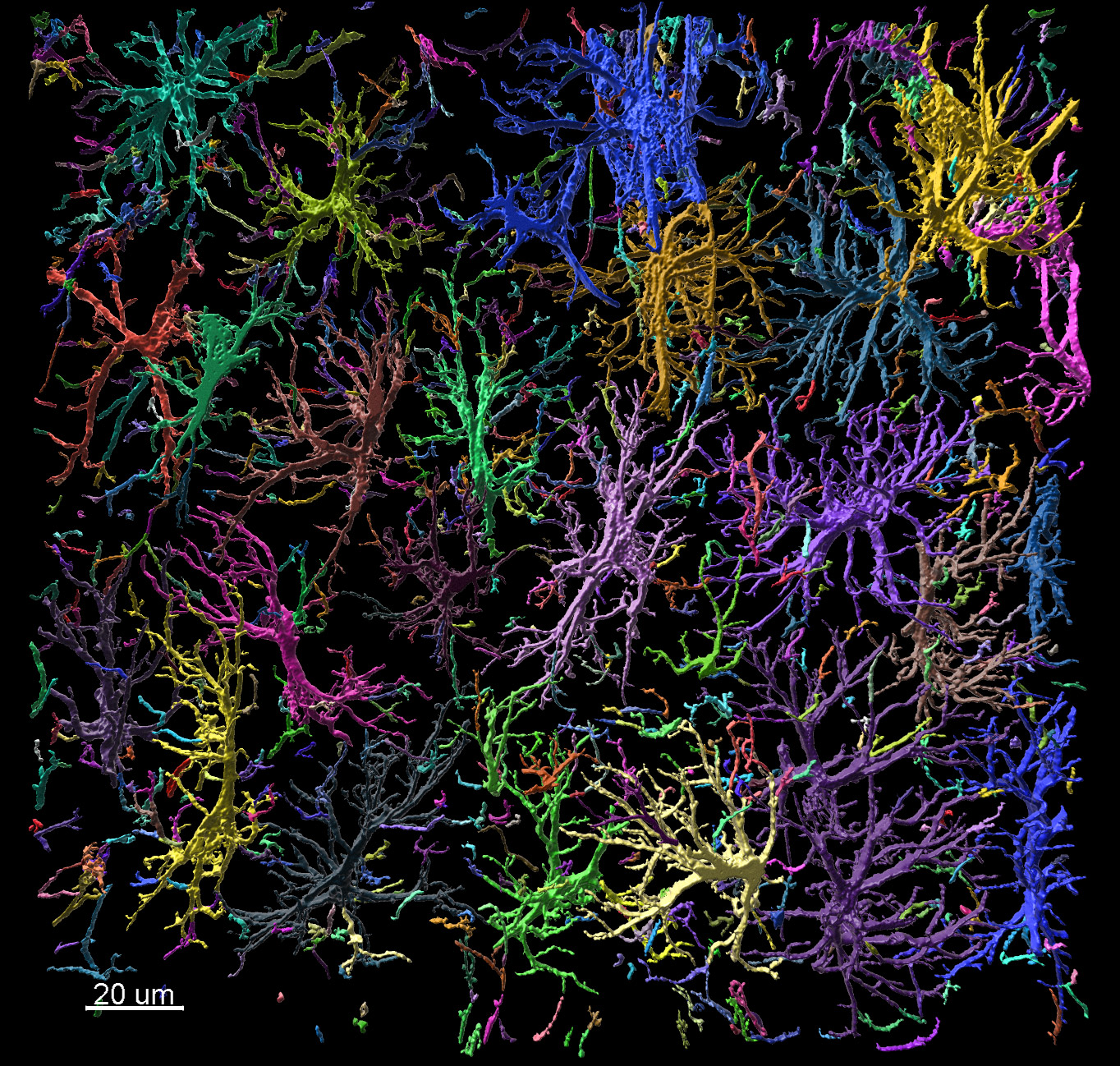
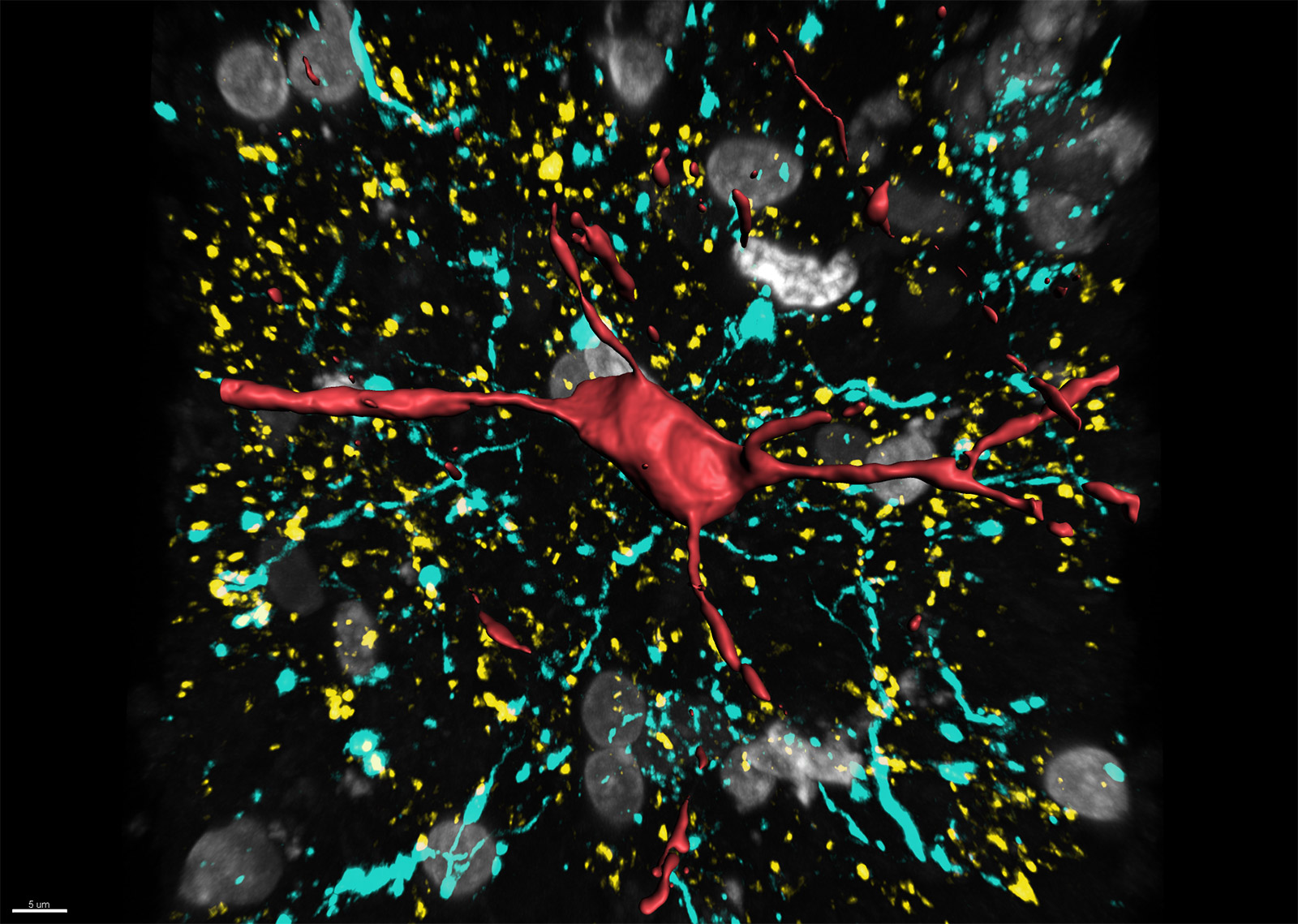
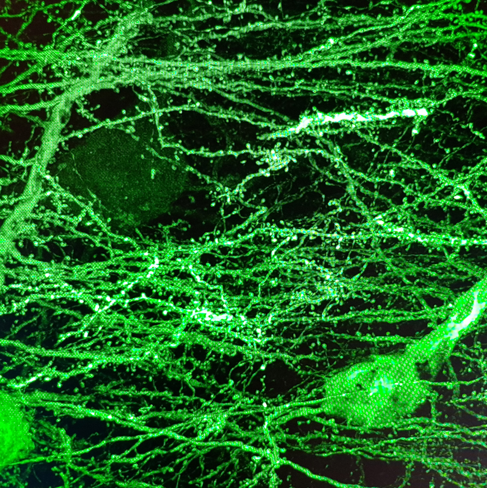
Neuroscience is a multidisciplinary branch of science focused on the study of the nervous system and how the brain works. The field studies nervous system functions, brain function and the related structures such as the spinal cord. It combines anatomy, physiology, cytology, molecular biology, developmental biology and modelling in order to understand neurons and neuronal circuits. As neuroscientists often balance on the cutting edge of science, they require sophisticated methods such as fluorescence labelling, optogenetics, photostimulation and state of the art image analysis. Learn about Andor and Imaris solutions for neuroscientists.

Applications and Techniques

Glia

For years glial cells were presumed to have housekeeping functions that only nourished, protected and swept up after the neurons, whose role seemed more obvious. Over the last couple of decades, research into glia (consisting of microglia, astrocytes, and oligodendrocyte lineage cells) has increased dramatically, which revealed their involvement in numerous supportive but essential functions, such as supplying in nutrients and oxygen, destroying pathogens and that they even play a role in neurotransmission. Although many aspects of these cells are well characterized nowadays, the functions of the different glial populations in the brain in health and disease remain unresolved.

In vivo intravital imaging or functional calcium imaging using a fast confocal spinning disc microscope such as Dragonfly is a crucial technique to monitor glial cells and neurons under various conditions. Optogenetics devices like Mosaic can be used to manipulate glia cell functions. Number, shape and other features of microglia can be analysed with 3D image analysis software – Imaris for Neuroscientists.

Contact our Application Specialists

Axonal Transport

Axonal transport is a cellular process responsible for the movement of cell organelles, vesicles, lipids and proteins to and from a neuron's soma, through the cytoplasm of its axon. Because of the axon length the transport cannot rely on diffusion, but it’s based on specialized motor proteins travelling along microtubules. Genetic mutations of those motor proteins dynein and kinesin are linked with some neurodevelopmental and neurodegenerative diseases.  Fluorescent labelling techniques have been crucial in enabling studying and visualization of axonal transport under physiological or pathological conditions in living neurons. This type of transport is a very fast event and fluorescence emission levels are often very low.

Therefore, fast and sensitive imaging such as Dragonfly equipped with sCMOS is crucial for studies of axonal transport in the living neurons. Time-lapse movies can be analysed using state of the art image analysis software Imaris for Cell Biologists or Imaris for Neuroscientists.

Contact our Application Specialists

Whole Brain Slices

Three dimensional visualization of intact brains or brain slices is a challenging but very desirable task in neurosciences. Application of tissue clearing and light microscopy have enabled investigating large volumes at micrometer resolution. Although light sheet microscopy gives advantage for examining full brain volumes, confocal microscopy is a preferred technique for acquiring cleared or native brain slices with higher resolution.

Imaging brain slices using Dragonfly spinning disk confocal equipped with a large FOV sCMOS camera and stitching microscopy tiles directly in Fusion (Imaris Stitcher algorithms) gives a perfect balance between the resolution and the size of the imaged sample. Imaris for Neuroscientists image analysis software does the rest: from data visualization, through counting to neuron tracing.

Contact our Applications Specialist

Optogenetics

Optogenetics is a biological technique, which allows for very precise control, modulation and activity monitoring of individual genetically modified neurons using light. The key players are neuronal ion channels, genetically modified with opsin genes, which act as “light sensors”. It is possible to use light to open the ion channels of a single neuron and fire the electrical signal. There are many supporting technologies that have helped enable this technique to become established: delivering specific wavelengths of light to opsin expressing cells with precise timing, controlling illumination through devices such as Mosaic, while sCMOS cameras are often the most suitable detector solution for these experiments, e.g. when high frame rates required for calcium imaging.

Optogenetics was applied in research involving formation of memories, addiction studies, providing better management of Parkinson’s disease tremors using deep brain stimulation or restoring vision.

Contact our Application Specialists

Expansion Microscopy

Instead of increasing the optical resolution of the microscope, Expansion Microscopy "expands" the sample isotopically. Different steps of the expansion protocol cause loss of the fluorescence signal so imaging expanded samples requires an instrument that is sensitive enough to detect low light signals. Other challenge is imaging a very large field of view and imaging deep into the sample. These requirements are problematic and difficult to achieve with conventional microscopes. Andor recommends the Dragonfly confocal equipped with either the iXon Ultra 888 back-illuminated EMCCD, or the Zyla 4.2P sCMOS cameras with optimised pinhole spacing for imaging deep into samples  and with Borealis for uniform illumination across the field of view.

Large data blocks can be stitched directly on the system using Imaris Stitcher algorithms and opened in Imaris for Neuroscientists for data visualization and analysis, including neuron and spine detection.

Contact our Application Specialists

Calcium Imaging

Calcium (Ca2+) is an important ion, which can be used as a rapid indicator of cellular activity from muscle cells (myocytes) to neurons and many others. Calcium Indicators (Ca2+ indicators) are continuing to provide great insights into fundamentals of cell biology, and for example how the cell responds during different disease states, or in response to therapeutic drugs. To make the most accurate determination of cell physiology the lowest illumination intensity and lowest possible concentrations of indicator dyes should be used. This inherently results in low photon emissions meaning a sensitive detector is highly important. There are two dominant camera technologies that are used for Ca2+ imaging experiments – sCMOS and EMCCD. In general, sCMOS cameras notably the Andor Zyla sCMOS camera have been the most widely used for Ca2+ imaging experiments. The new

Sona back-illuminated series builds on the performance of the Zyla models keeping the important high speeds, high resolution and class leading quantitative accuracy.

Contact our Application Specialists

Products

Learning Resources