Resources
 Part of the Oxford Instruments Group
Expand
Collapse
Part of the Oxford Instruments Group
Andor’s product portfolio for virology provides a range of imaging solutions that are helping to further our understanding of viruses. This includes addressing questions such as how viruses are transported within the cell, how viruses hijack the cell for replication, and how they interact with, and evade the immune system. Other areas include development of techniques to detect viruses in the environment, and in the development of potential anti-viral treatments.
Our portfolio includes the most sensitive imaging cameras to detect fluorescently labelled viruses, high-speed confocal imaging systems that allow multiple imaging techniques, and powerful software to help unlock the complexities of virus to host cell interactions.
Request PricingLearn how different research groups have been using iXon EMCCD cameras to help understand different aspects of the virus cycle.
Fluorescence imaging techniques for study of inactivated viruses, or for live tracking experiments demand the most sensitive detectors. From Adenovirus to Zika, iXon EMCCD cameras have a proven history as the detectors of choice for the most challenging virology imaging applications. Andor’s latest iXon Ultra and Life models are the most sensitive EMCCD cameras available.
The latest back-illuminated sensor sCMOS technology narrows the gap in ultimate sensitivity to EMCCD making these cameras more applicable to a wider range of virology studies than previous sCMOS cameras. Andor’s new Sona back-illuminated sCMOS series fully exploits the sensor technology providing high sensitivity, and exceptional speeds over the widest possible fields of view.
EMCCD and back-illuminated cameras are broadly suitable for the main techniques used for imaging viruses – e.g. confocal, TIRF, single molecule localization super resolution or FRET. The technologies each have their own benefits – so which one will suit best? The following guide provides a comparison of the different camera models against various experiment requirements
| Back-illuminated EMCCD cameras | Back-illuminated sCMOS cameras | |||
| Application Requirement | iXon Life/ Ultra 888 | iXon Life/ Ultra 897 | Sona 4.2B-6 | Sona 4.2B-11 |
| Capture weakest signals | ||||
| High Sensor Resolution1 | ||||
| Image a Wide Field of View | ||||
| High speed Imaging2 | ||||
| High Dynamic Range | ||||
| Long exposure suitability3 | ||||
| High quantitative accuracy | ||||
| Summary | When ultimate sensitivity is required. The largest field of view and fastest EMCCD option. | When ultimate sensitivity is required and wide field of view is not a priority. | Highly flexible imaging solution provides high sensitivity, field of view and highest speeds. | For a balance of high sensitivity and field of view. |
1 Effective image resolution for light microscopy is set by the microscope objective (Resolution=λ/2NA) and the technique used. Smaller pixel sizes will meet, or exceed Nyquist sampling at lower objective magnifications and may help with localization based super resolution or deconvolution.
2 EMCCD cameras may run at high frame rates by cropping the sensor to a smaller field of view.
3 sCMOS cameras are most suitable for short exposures. EMCCD cameras can be used for extended exposures into minutes due to exceptionally low dark current.
Still not sure which camera to choose, contact our application specialists.
In addition to the world’s most sensitive virology cameras, we deliver the best-integrated solution for image acquisition and analysis, combining Dragonfly high speed confocal for acquisition with Imaris software for imaging for tracking, statistical analysis and 3D rendering.
The Dragonfly allows a number of imaging modalities that are used for virology studies such as confocal spinning disk, TIRF, dSTORM and SRRF-Stream+. All these imaging modalities are delivered in one single system, allowing the user to select the best match for each specific experiment. Find out more about how the new Dragonfly system is being used in virology in our solution note: How to use confocal microscopy to image viruses.
Request Pricing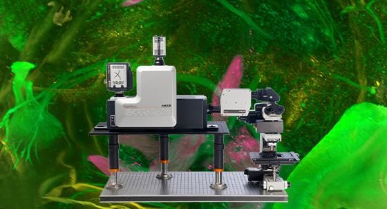
With the latest developments in labelling viruses and new techniques we are gaining further insight into the complexities of how viruses interact with host cells such as virus transport and assembly. The interplay between virus, the host cell and how the immune system seeks to counteract viruses can be greatly aided by effective analysis software. Imaris provides a range of functionalities that can empower your research. To find out more about how Imaris is being used in virology studies such as CoV-2 research please view our solution note.
Request Pricing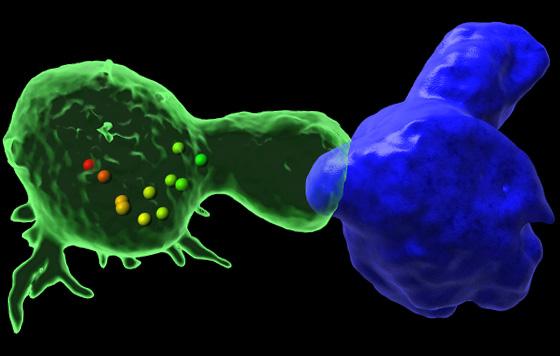
Please take a look below at some of the virology articles available in the learning center. You can also listen to our virus podcast here.
Advances in light microscopy techniques and labelling approaches have allowed for much greater flexibility in terms of experimental capabilities within virology research. Super resolution techniques such as STORM, PALM, SIM, TIRF and SRRF-Stream+ allow the diffraction limit of light to be overcome and improve imaging resolution down to 50 nm, or beyond.
Improved resolution affords greater insight into the intricate and dynamic nature of how viruses interact with the host cell during the infection cycle. The range of what can be studied using light microscopy techniques can therefore span from studying the general histopathological effects within tissue samples, detection of virus within a sample, localization of viruses within cells and potentially, tracking of live viruses during the virus infection cycle.


© Oxford Instruments 2026