Resources
 Part of the Oxford Instruments Group
Expand
Collapse
Part of the Oxford Instruments Group
Application Notes
Author: Sachin Verlekar, Christophe Galland, Institute of Physics and Center of Quantum Science and Engineering, Ecole Polytechnique Fédérale de Lausanne (EPFL), 1015 Lausanne, Switzerland
Published: 01 Jun 2025 · Last updated: 24 Jun 2025
The light emitted by individual molecules - in terms of intensity as well as spectra - can be controlled via their coupling to plasmonic nanocavities that change the local electromagnetic environment. Using DNA origami nanotechnology, we precisely position single organic fluorophores in the hotspot of plasmonic nanocavities. Careful design of the nanodimer-on-mirror (NDoM) nanocavities allows for cavity-mediated fluorescence reshaping, leading to far detuned (> 100x the bare emitter linewidth), strong emission in the near-infrared region.
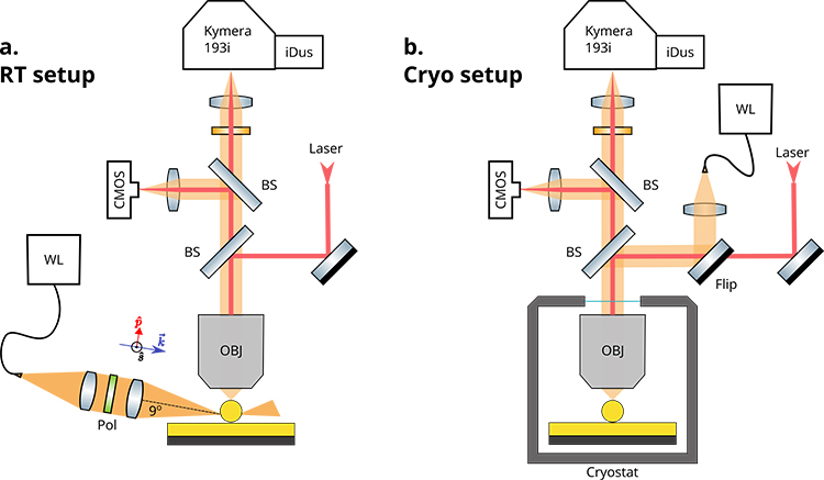
Fig. 1 Experimental Setup: Schematic representations of experimental setups used for (a) darkfield scattering and (b) cryogenic fluorescence microscopy of single nanocavities.
Two home-built experimental setups are used for this study. The plasmonic modes of individual nanocavities are characterized via dark-field scattering spectroscopy where white light impinging from a high angle (81 degrees w.r.t the vertical, Fig. 1a) is scattered by the nanocavities into a microscope objective. The nanocavities are then measured in a different setup (Fig. 1b) at cryogenic temperature (4K) where fluorescence spectra are acquired using CW laser excitation. In both experimental setups, the spectra are measured by aligning the collected signal into a spectrograph (Andor Kymera 193i) equipped with a CCD (Andor iDus). Their compact footprint as well as high sensitivity across the VIS-NIR spectral range allows for easy integration into both setups.
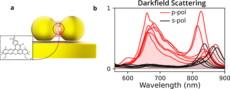
Fig. 2 Darkfield Scattering: Individual NDoM nanocavities, as shown schematically in a are characterized via polarization-resolved darkfield scattering spectroscopy. b shows experimental spectra from a few nanocavities.
We use DNA origami to place a single ATTO 590 fluorophore precisely between two gold nanospheres (60 nm diameter), forming a nanodimer (ND) cavity with a gap size of 4-5 nm. NDs are then deposited on either glass (nanodimer on glass, NDoG) or ultra flat Au films (nanodimer on mirror, NDoM, see Fig. 2a). The plasmonic modes of single nanocavities are investigated via dark-field scattering spectroscopy. Figure 2b shows darkfield scattering spectra of NDoMs, which reveal resonances in the visible (630-700 nm) as well as the NIR region (> 800 nm). The NIR resonance arises from the interaction between the bonding dipolar mode of the ND and the mirror.
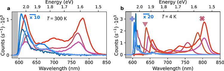
Fig. 3 Spectral Reshaping: Single molecule fluorescence is reshaped using NDoM cavities at ambient (a) as well as cryogenic (b) temperatures. Shades of blue indicate fluorescence of uncoupled dyes while the shades of red represent results from NDoM cavities.
As shown in Fig. 3, the dual mode nature of NDoM cavities allows for spectral reshaping of single fluorophores due to a high Purcell factor (up to ~105) and coupling to the highly radiative NIR mode. In addition to an enhanced spectral intensity, a dramatic reshaping of the emission spectrum is seen, characterized by a broadened zero-phonon line as well as the appearance of a distinct new emission peak around 750-800 nm. We confirm that the NIR emission inherits the peak wavelength and linewidth of the plasmonic mode.
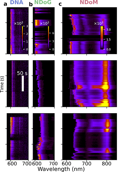
Fig. 4 Single molecule fluctuations: Typical timeseries of spectra are shown, with each column containing three examples of bare fluorophore in DNA (a), fluorophore in NDoG (b) and fluorophore in NDoM (c). A cw laser at 590 nm is used to excite the fluorophore and the timeseries is collected at a frequency of 1 Hz.
The kinetic mode of Andor iDus allows us to capture time series of the full spectra, allowing for the observation of dynamic single-molecule events. Examples of such time traces are shown in Fig. 4 for all the configurations studied. Sharp ZPLs, which blink and bleach, are seen for individual fluorophores, while a lot more spectral broadening and wandering of the ZPL is seen in the case of the NDoG. In the NDoM, this broadening allows for the coupling to the NIR mode. When the ZPL wanders closer to the NIR mode, the intensity of the NIR mode clearly increases, hence indicating a change in the coupling and overall reshaped spectra.
In this work, we have demonstrated that single molecule fluorescence can be dramatically reshaped through coupling with DNA-origami-positioned plasmonic nanocavities, specifically nanodimers on mirror (NDoM) configurations. By precisely engineering the local electromagnetic environment, we observe coupling between the zero-phonon line (ZPL) of the fluorophore and the plasmonic modes of the cavity, leading to enhanced emission in the near-infrared (NIR) region - far beyond the natural spectral profile of the emitter.
Verlekar, S., Sanz-Paz, M., Zapata-Herrera, M., Pilo-Pais, M., Kołątaj, K., Esteban, R., Aizpurua, J., Acuna, G.P. and Galland, C. “Giant Purcell broadening and Lamb shift for DNA-assembled near-infrared quantum emitters”. ACS Nano 2025, 19, 3, 3172–3184 https://pubs.acs.org/doi/abs/10.1021/acsnano.4c09829 .
European Union’s Horizon 2020 research and innovation program (Grant Agreement No. 820196, ERC CoG QTONE), and Swiss National Science Foundation (project number 214993)

© Oxford Instruments 2026
