Resources
 Part of the Oxford Instruments Group
Expand
Collapse
Part of the Oxford Instruments Group
X-ray and neutron imaging applications such as material science & industrial inspection, rely on the ability to capture X-ray and Neutron images with the utmost precision and accuracy.
Andor's extensive camera portfolio of sCMOS, CCDs, ICCD and EMCCDs offer a dynamic range of solutions to address challenges in tomography, ptychography and imaging applications both in situ & ex situ. Andor offers a portfolio of cameras for both soft and hard X-ray & neutron imaging applications including direct detection CCDs & sCMOS (X-ray) and indirect fibre and lens coupled cameras (X-ray & Neutron).
X-ray and neutron imaging or Radiography is an imaging technique in which X-rays or Neutrons are used to probe the internal composition and structures of materials. X-rays and Neutrons are absorbed by samples depending on the density and elemental mass. In the case of X-rays the attenuation of X-rays through a material is linked to a materials density whereas the attenuation of neutrons through a material is linked to the elements present in a material as neutrons only interact with atomic nuclei. Due to the difference in material interactions often X-ray and neutron imaging techniques are used in conjunction to provide a complimentary non-destructive insight into the structure of materials. Key applications of radiography include the inspection of materials (e.g. steels, ceramics and alloys), biological samples (e.g. cells, plants), forensic and geological samples (e.g. rock samples and meteorites).
X-ray images can be acquired either through direct absorption of X-rays onto a 2D silicon based camera sensors for energies <20 keV or though using scintillators lens coupled to a CCD or sCMOS detector at higher energies such as >20 keV. Neutron images are typically acquired using a scintillator (generally made of Lithium-based materials (e.g. LiF(ZnS) or plastic)) with a CCD or sCMOS detector offset at 90 degrees with mirrors to protect the sensor apparatus from the damaging neutron beam.
Andor offers a comprehensive range of cameras ideal for direct X-ray imaging and in-direct X-ray and neutron imaging applications. To learn more see here.
Contact Application SpecialistsX-ray Computed Tomography or X-ray CT is a non-destructive technique that be used to study the internal structure and composition of materials using the absorption or phase contrast changes that occur then X-rays pass through a material. X-ray CT works through the acquisition of a series of single 2D images of a revolving 3D sample which are then combined to render a 3D image of a sample. The absorption/attenuation of X-rays through the sample give rise to changes in contrast which is determined by the density of the different components within a sample and their individual to absorb or phase shift X-rays. X-ray CT is used widely in the study of materials (e.g. metals, glasses, ceramics), art (e.g. scrolls, paintings, statues), energy storage devices (e.g. batteries) and biological systems (e.g. cells within the water window of the X-ray spectrum or plant structures).
Andor offers a comprehensive range of both slow scan CCD cameras direct soft X-ray/EUV tomography and indirect hard X-ray tomography applications and fast sCMOS cameras for direct soft X-ray/EUV tomography and indirect hard X-ray tomography applications. To learn more see here.
Contact Application SpecialistsNeutron Tomography is a non-destructive technique that can be used in the study hydrogenous fluid dynamics or fluid distribution (e.g. water) in metal objects, corrosion processes, complex archaeological artefacts or geological samples inner structures. It can also be used for quality control of engineering systems (e.g. combustion engines, Li-ion batteries, fuel cells), to discriminate isotopes of the same element or to study water transport in biological materials (e.g. plants).
Neutron images are typically acquired using a scintillator (generally made of Lithium-based materials (e.g. LiF(ZnS) or plastic)), lens – coupled to a CCD or sCMOS detector. Andor offers both a comprehensive range of large area slow scan high sensitivity CCD cameras idea for weak signal long exposure acquisitions and large area, low noise high frame rate sCMOS cameras ideally suited to fast tomographic applications. To learn more see here.
Contact Application SpecialistsIn situ X-ray & neutron imaging & tomography can be used to study a wide range of transient phenomena in material science, biology and physics such as the melt and crystallisation of metals, tensile testing of materials and absorption of water into biological systems such as root systems.
In situ imaging and tomography can be demanding in terms of data acquisition times and frame rate requirements for scientific cameras. Fast transient phenomena imaging requires high frame rates and when combined with tomography requires even high frame rates. Additionally, to acquire images rapidly requires a high sensitivity camera with low noise readout to reach the required signal to noise level for each individual frame and minimise total acquisition time. A highly sensitive, low noise and fast camera critical to make the most out of both high flux and low flux in situ environments.
Andor offers a comprehensive range of both slow scan CCD cameras for direct soft X-ray/EUV in situ applications and indirect hard X-ray in situ applications. Additionally, Andor offers a market leading range of fast sCMOS cameras for direct soft X-ray/EUV in situ applications and indirect hard X-ray in situ applications. To learn more, see here.
Contact Application SpecialistsPtychography is a method to reconstruct the crystal structure (image) of a specimen from the diffraction patterns obtained from each point (area) scanned over a specimen using a convergent probe so that a part of the illuminated area overlaps. “Ptycho” means “fold” in Greek.
X-ray ptychography has been shown to be the most robust scanning method of coherent diffraction imaging (CDI) and has the advantage of being a lensless technique so avoids the difficulties associated with producing X-ray lens optics. Once a ptychography pattern is obtained through scanning X-rays through a sample an iterative feedback algorithm is used to reconstruct an image of the sample. X-ray ptychography provides high spatiotemporal resolution for probing dynamic phenomena in both 2D and 3D.
X-ray ptychography is used increasingly commonly in material science (i.e. the study of metals, alloys, composites, ceramics, and 2D materials), semiconductor fabrication and quality inspection, medical, geological, archaeological and engineering.
Andor has an excellent array of deep depletion direct detection CCD cameras well suited to soft X-ray/EUV ptychography <20 keV and has recently brought out a range of cutting edge direct detection sCMOS cameras for direct EUV and soft X-ray ptychography applications. To learn more, see here.
Contact Application SpecialistsAndor offers a comprehensive portfolio of open fronted direct X-ray detection cameras (SO), and high throughput fibreoptic and lens coupled indirect detection solutions to meet every need.
Andor incorporates the latest in direct detection sensor technologies into its extensive product portfolio.
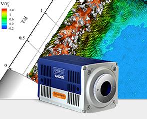
We offer cutting edge direct detection sCMOS cameras capable of directly detecting X-ray/EUV photons are market leading frame rates (up to 74 fps at 4.2 megapixel resolution!). Direct detection sCMOS is ideally suited to fast imaging, tomography, ptychography, lithography and spectroscopy applications at <5 keV energy ranges. To see our range of direct detection sCMOS cameras see here.
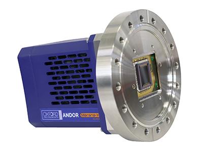
For long exposure, 10 eV – 20 keV energies and large area applications Andor also offers a comprehensive range of ultra-sensitive open fronted direct detection CCD cameras deep cooled to -100 ゚C enabling relatively long camera exposures over large fields of view (up to 61 x 61 mm!). Andor’s range of direct detection CCD cameras are ideal for applications such as resonant inelastic X-ray scattering (RIXS), imaging, tomography, ptychography To see our full range of direct detection CCD cameras see here.
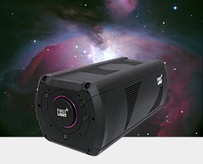
For hard X-ray (>15 keV) & neutron applications such as imaging, tomography and in situ studies it can be critical to make the most of each incident photon or neutron into your setup. Fibre coupled cameras offer a compact, versatile, high throughput solution for X-ray and neutron imaging applications.
Fibre coupled cameras can be equipped with a variety of scintillators tailored to the application and energy range. Light from the scintillator is efficiently channelled down the fibre couple into the camera sensor to provide a high resolution, high efficiency image. Fibre cameras enable the maximum throughput through an imaging system and in turn this enables the end user to make the most out of their high speed camera to operate at the maximum frame rate possible or make the most of every incident X-ray photon in flux limited systems.
Andor offers two fibre coupled camera solutions for imaging:
For hard X-ray (>15 keV) & neutron applications such as imaging, tomography and in situ studies lens coupled cameras offer an effective versatile solution to X-ray and Neutron imaging.
Lens coupling offers several advantages for X-ray and neutron imaging:
Andor offers an extensive portfolio of cameras for a diverse array of neutron and X-ray lens coupled experimental setups including large area, low noise, fast sCMOS cameras for rapid techniques such as high-resolution tomography and in situ imaging learn more here. Additionally, Andor’s comprehensive range of CCD cameras offer deep cooled, low noise and high dynamic range performance ideal for low flux imaging & tomography applications here.

© Oxford Instruments 2026