Resources
 Part of the Oxford Instruments Group
Expand
Collapse
 Part of the Oxford Instruments Group
S. cerevisiae is one of the long-time classic model organisms that remains highly relevant today. As one of the simplest eukaryotes (containing membrane bound organelles), and indeed the first eukaryotic organism to be sequenced with a genome size of ~12 Mbp, it can be used for studies of common pathways in higher organisms such as humans. [1][2] In addition to important applications within the food industry and biotechnology areas.
As yeast share the same cell division processes with humans, S. cerevisiae is well established for studies of cell division and cancer research applications. Yeasts have simple nutritional requirements and can be easily grown in standard lab conditions. Recently, s. cerevisiae has featured in important research into neurodegenerative studies such as Parkinson’s disease. Accumulation of the protein alpha-synuclein over time is a key factor of the disease; yeast are similarly affected by accumulation of this protein making them an ideal model to study the development pathways involved, and for screening of potential drug treatments.
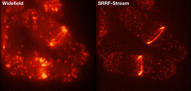
On imaging yeast cells one must consider that they tend to have low levels of fluorescence making a high sensitivity camera such as the Andor iXon EMCCD or back illuminated Sona essential. Super-resolution techniques such as Andor’s SRRF-Stream (shown in Figure 2) can also be particularly effective as it is possible to image the fine structure and processes within these cells in real time.

Figure 2: A frame from comparative 3D projection montages of fission yeast lifeAct expressing strain. Recorded with standard widefield versus SRRF-Stream widefield, using identical exposure times the improved resolution can readily be observed. Strain courtesy of Mohan Balasubramanian’s laboratory (U. Warwick) Sample courtesy of Gautam Dey (Buzz Baum laboratory at UCL).
References
Learn More from the Model Organism Series
Date: February 2019
Author: Dr Alan Mullan & Dr Aleksandra Marsh
Category: Application Note
