Resources
 Part of the Oxford Instruments Group
Expand
Collapse
Part of the Oxford Instruments Group
Application Notes
Author: Dr Alan Mullan & Dr Aleksandra Marsh
Published: 01 Feb 2019 · Last updated: 31 Jul 2025
C.elegans is a nematode worm and is significantly anatomically simpler than a human, however, it does share many similarities at the molecular level making it a good candidate for a model organism.
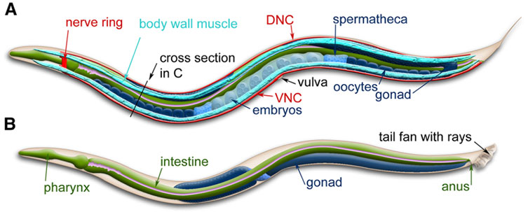
Figure 10: C. elegans anatomy. Major anatomical features of a hermaphrodite (A) and male (B) viewed laterally. Modified from Corsi et al.
As with most of the other model organisms considered here C. elegans is easy to look after, feed and maintain throughout their 2-week lifespan. C. elegans has few discernible external features, and as such, initially appears a poor subject for study. However, it does have many advantages: its transparency makes it possible to observe the fate of individual cells using simple microscopy, or more advanced techniques such as SPIM or Light Sheet Microscopy. C. elegans grown in large numbers, can be easily screened for effects of novel drugs on complex processes involved in human disease. C. elegans is particularly useful the study of ageing processes because the organism passes through several distinct phases of life which can be observed physiologically and genetically. Moreover, C. elegans is also used to study neural development; it is well suited for this application due to the availability of a comprehensive connectivity map and only 302 neurons and ~7000 synapses.
Imaging of C. elegans using SPIM, Light Sheet Microscopy or Spinning Disk Confocal requires fast live-cell imaging at high resolution; Andor recommends the Zyla 4.2 Plus and Sona sCMOS cameras for these applications. Zyla sCMOS affords high resolution imaging performance and Sona is the most sensitive sCMOS camera with a very large field of view, superior longevity and quantitative accuracy.
References
Learn More from the Model Organism Series

© Oxford Instruments 2026
