Resources
 Part of the Oxford Instruments Group
Expand
Collapse
Part of the Oxford Instruments Group
Application Notes
Author: Dr Alan Mullan & Dr Aleksandra Marsh
Published: 01 Feb 2019 · Last updated: 19 Aug 2025
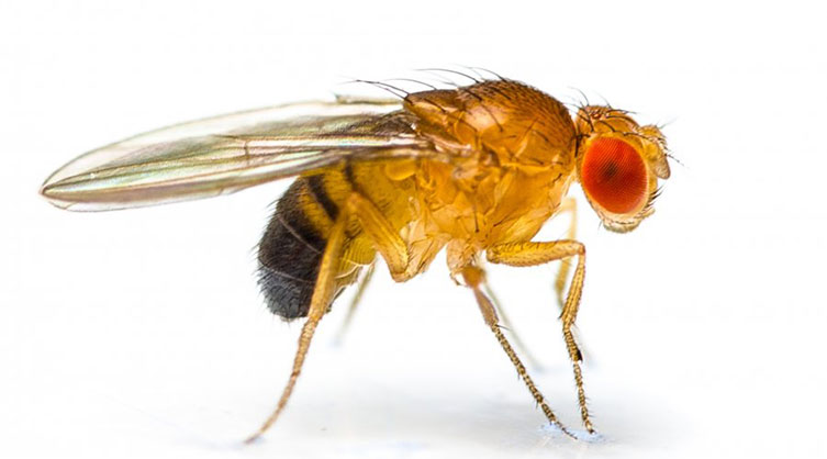
Figure 6: Drosophila melanogaster (image credit Shutterstock)
Wild D. melanogaster or fruit flies are originally associated with the Marula fruit indigenous to South Africa.[1] In the laboratory, the fruit fly has been a key model organism since the very first studies of genetics. It was the humble fruit fly that provided us with information on genetic inheritance of chromosomes at the phenotype level.
The fruit fly is simple to work with, with a relatively short lifecycle/generation time of 12 days and its small size allows it to be produced in large numbers. These practical considerations make it suitable for many studies. The fruit fly is well understood at the phenotype level and has a simple genome, enabling molecular genetics studies. Despite this, it still has ~60% of the genes involved in human genetic diseases and some cancers.
The fruit fly has also been used as a model for more complex studies, such as development and processes involved in cognitive behaviour, memory and learning studies. Recently it has been used in studies on wound repair and infection prevention. [2] Imaging of D. melanogaster can be done using light sheet microscopy to investigate embryo development. [3] Andor have developed cameras specifically designed to serve as detectors for high-speed light sheet microscopy; the Sona and Zyla range of sCMOS cameras offer a large field of view and high resolution without compromising read noise or frame rate.

Figure 7: Egg-chamber from Drosophila melanogaster at stage 10 captured with a Zyla 5.5 camera. Visible is the large oocyte with its tiny meiotic nucleus. Stained are: DAPI (blue) to show the DNA, WGA-657 (green) to show membranes, primarily visible are nuclear membranes. Also stained by fluorescent in situ hybridization is the oskar mRNA that is enriched at the posterior cortex. Detection: Cy3-coupled tyramide amplification. Captured using 25x objective with 0.8 numerical aperture. Courtesy of Dr Helena Jambor, Tomancak Lab at MPI Dresden.
References:
Learn More from the Model Organism Series

© Oxford Instruments 2026
