Resources
 Part of the Oxford Instruments Group
Expand
Collapse
Part of the Oxford Instruments Group
Application Notes
Author: Dr Alan Mullan
Published: 01 Feb 2019 · Last updated: 19 Aug 2025
Zebrafish (Danio rerio) are small tropical fish (2.5-4 cm long) found natively in south Asia. First used in the 1960s in biological research, Zebrafish have proven to be excellent model organisms for developmental and disease studies. They are now very well characterised genetically with genome size ~1.5 Gbp. Zebrafish can be grown easily, maintained in lab conditions and produce young all year round.
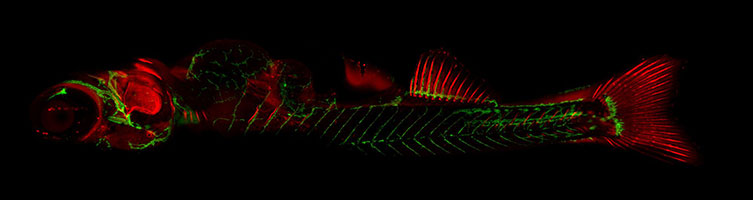
Figure 13: 30 days old transgenic zebrafish (flk:EGFP, lyve1:dsRED), flk positive cell are from the endothelial lineage and give good visualization of the blood vessels. lyve is a marker for lymphatic endothelial cells and gives good visualization of the lymphatic vessels. The fish was anaesthetized and mounted in 0.5% agarose on a glass bottom plate. Stitched image of 6 x 3 tiles. 25x Water objective. Courtesy of Dr. Karina Yaniv, Or Nelkenbaum, Prof. Lia Addadi, Weizmann Institute.
Zebrafish are commonly used to the study the early stages of development. Moreover, they are well suited for embryonic studies because of their relatively transparent body during the larval development stage. This provides excellent optical clarity, making them a practical choice for easy observation. In contrast, the early stages of development are harder to study in mice since they are in utero. Modern cameras such as the Sona 4.2B-11 offer very wide fields of view and this means that whole zebrafish embryos can be imaged in a single frame.
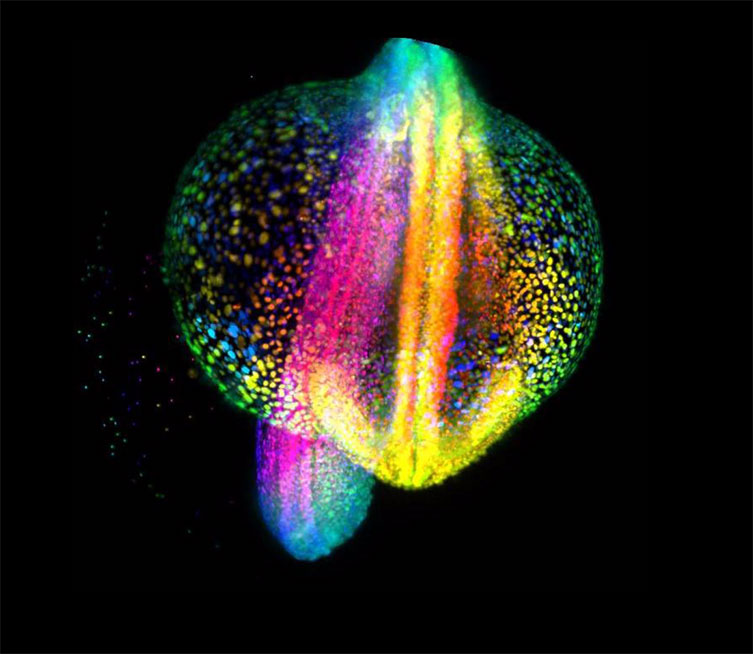
Figure 14: Image courtesy of Gopi Shah, Max Planck Institute of Molecular Cell Biology and Genetics, Dresden. Light-sheet fluorescence microscopy imaging of Zebrafish embryo gastrulation from 4 to 18 hours post-fertilization, where each cell nucleus is labelled with GFP. Cells are color-coded for depth to visualize how dynamic cell reorganization gives rise to the body axis of zebrafish
Zebrafish, unlike C.elegans, possess most of the major organ systems of vertebrates like humans, for example, heart, eyes and kidneys, therefore, organ development can be studied. Features such as blood vessels can also be seen at relatively low magnification. Martin et al. have recently demonstrated that the derivation of heart rate from multiple zebrafish can be done using a high-throughput platform implementing brightfield microscopy using Andor’s Zyla 4.2 sCMOS camera. [1]
Approximately 80 % of the genes involved in human diseases have an equivalent in Zebrafish. Due to good characterisation of zebrafish genes, it is possible to target specific genes, turn them on or off, or introduce foreign genes; this makes zebrafish very versatile for modern research.
Read more about Imaging Thick Samples of Large Model Organisms such as Zebrafish, Drosophila and C.elegans in our full solution note.
References
Learn More from the Model Organism Series

© Oxford Instruments 2026
