Resources
 Part of the Oxford Instruments Group
Expand
Collapse
 Part of the Oxford Instruments Group
Many microscopy techniques have been developed in life sciences to study biological organisms and their underlying biochemical and molecular processes. Therefore, life science research can span many orders of magnitude in scale, from studying single molecules through to whole model organisms.

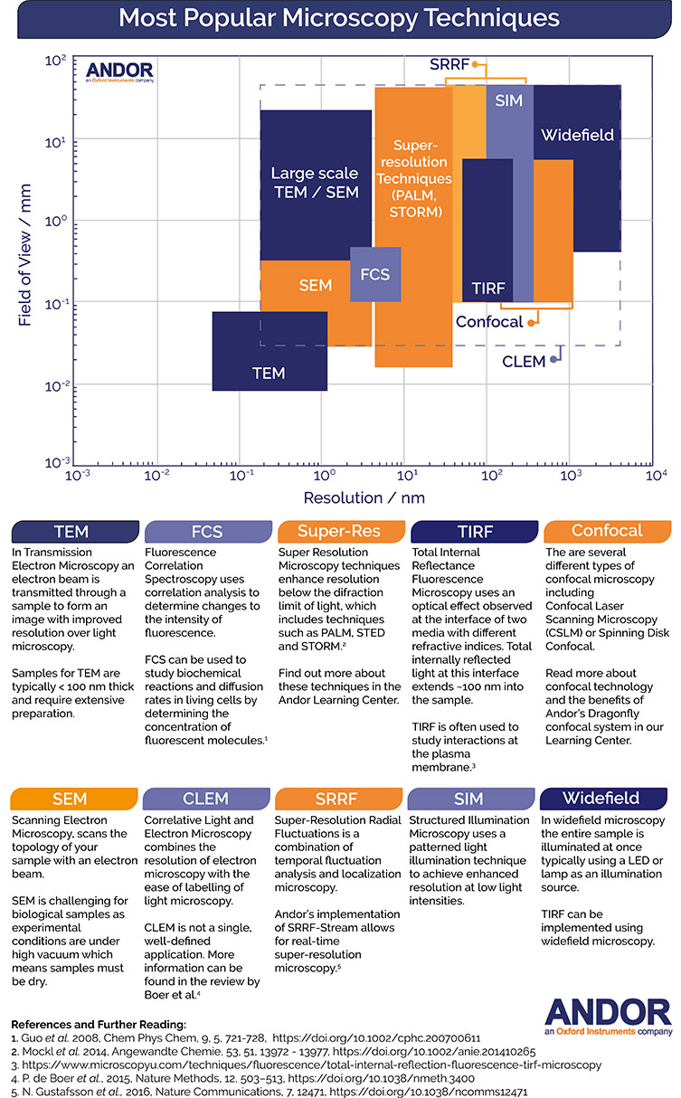
A wide range of microscopy techniques are available for life science research. The figure below provides a summary of the range of experimental methods available, shown as a function of their field of view and lateral resolution. Please see the table below for a detailed description of acronyms used in field of microscopy.


In the table below some of the common acronyms and their full names are detailed. Links to articles in our learning centre provide a more in-depth description of the techniques and their applications in life science research. It should be noted that CLEM is a mix of various electron and light microscopy techniques, not a single, well-defined application. More information can be found in the review by Boer et al.
| Common acronym | Full name |
| CLEM | Correlative Light and Electron Microscopy |
| CLSM | Confocal Laser Scanning Microscopy |
| FCS | Fluorescence Correlation Spectroscopy |
| FRET | Fluorescence Resonance Energy Transfer Microscopy |
| LSFM | Light Sheet Fluorescence Microscopy |
| PALM | Photoactivated Localization Microscopy |
| SDCLM | Spinning Disk Confocal Laser Microscopy |
| SEM | Scanning Electron Microscopy |
| SIM | Structure Illumination Microscopy |
| SPIM | Selective Plane Illumination Microscopy |
| SRRF | Super-Resolution Radial Fluctuations |
| STED | Stimulated Emission Depletion |
| STORM | Stochastic Optical Reconstruction Microscopy |
| TEM | Transmission Electron Microscopy |
| TIRF | Total Internal Reflectance Fluorescence Microscopy |
1. Guo et al. 2008, Chem Phys Chem, 9, 5, 721-728, https://doi.org/10.1002/cphc.200700611
2. Mockl et al. 2014, Angewandte Chemie, 53, 51, 13972 - 13977, https://doi.org/10.1002/anie.201410265
3. https://www.microscopyu.com/techniques/fluorescence/total-internal-reflection-fluorescence-tirf-microscopy
4. P. de Boer et al., 2015, Nature Methods, 12, 503–513, https://doi.org/10.1038/nmeth.3400
5. N. Gustafsson et al., 2016, Nature Communications, 7, 12471, https://doi.org/10.1038/ncomms12471
