Resources
 Part of the Oxford Instruments Group
Expand
Collapse
Part of the Oxford Instruments Group
Application Notes
Author: Dr Alan Mullan & Dr Jo Walters
Published: 01 Feb 2023 · Last updated: 21 Feb 2023
Cell lines are very effective research models as they represent the specific types of cells we may be interested in. However, in a complete multicellular organism, cells do not exist in homogenous suspensions, or monolayers in multicellular organisms. In a real organism, there are many specialised cell types that perform specific and very different functions – a cardiomyocyte in the heart is very different to a mast cell. Cells are also part of a connected network with other cell types in specific spatial organisations and this is known to change their behaviour e.g. response to different drugs, or impact specific cell types within a tissue differently.
The easiest way to start to study tissues is by using fixed tissue samples, but since this is a fixed 2D slice or set of slices this will have limitations. A true living 3D tissue as a research model is more difficult to access and observe. Firstly because of access to suitable cell tissue samples. Secondly with problems in imaging these relatively thick samples. Read more about how to image thick tissue samples.
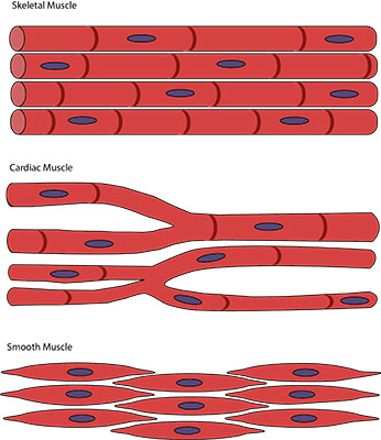
Skeletal muscle cells are organised in bundles of long cells that contract under voluntary control (conscious control via the somatic nervous system), and permit motor force and movement. Muscle cells, also called myocytes, contain multiple nuclei distributed through each fibre which is related to their need to produce many proteins and enzymes to function. Muscle fibres may be arranged in different organisations to allow a range of muscle types that allow control of motion of different joints.
Cardiac muscle cells have a number of differences that relate to their specialised role in pumping blood around the body such as involuntary action and a higher abundance of mitochondria providing a much greater resistance to fatigue. Cardiac muscle tissue has a high oxygen demand and deprivation of Oxygen will lead to death of muscle cells.
A third group of muscle tissue is smooth muscle. Smooth muscle cells are highly elastic fusiform (spindle-shaped) cells organised in sheets, with a primary role of contraction. Smooth muscle is found widely in the body playing vital roles in the digestive system, the reproductive and renal systems, respiratory and cardiovascular systems.
The different types of muscle cells thus comprise a number of highly specialised cells with important functions in the body, making their study of significance for understanding of disease states and their treatment. For cardiac cells and blood flow, high speed sCMOS cameras like ZL41 Cell and Sona-6 may be used to image at speeds of 100-500 fps.
Connective tissues are found throughout the body between other tissue types and organs. Connective tissue proper typically consists of three major components. These are:
1. A fibrous component such as elastic and collagen fibres that provide strength to the tissue
2. A ground substance, a gelatinous material containing glycoproteins, proteoglycans and many other biomolecules and water of the extracellular matrix
3. Cells – which include adipose, fibroblasts and macrophages according to the type of connective tissue
There are many different types of connective tissue from the highly dense, fibrous collagen rich tissues of ligaments to loose adipose tissue composed of lipid rich lipocytes. Blood, cartilage and bone are also classified as special connective tissues.
There are many disorders that are related to connective tissues. The most common example of a connective tissue disease is rheumatoid arthritis, which is an inflammation of the membranes surrounding the joints results from an autoimmune response to these tissues causing progressive damage.
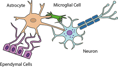
Nervous tissue studies are one of the most intensely researched due to the role nervous tissues play in neurodegenerative diseases. Nerve tissues of the brain, spinal cord and nerves are highly complex compared to more simple tissue types like sheets or bundles of muscle or connective tissues. Nervous tissue is composed of neuronal and a number of supporting types of neuroglia cells such as astrocytes and microglia. Neurons are polarised cells and it is these cells that allows the flow of electrical signals along their membrane and transmission of signals over distance. The dynamics of signalling within these networks that are often what must be studied making it demanding from an imaging perspective. The development of optically active fluorescent labels and photostimulation tools such as Andor’s Mosaic and MicroPoint alongside visualisation and analysis software like Imaris have greatly aided study of nervous tissues and the complexities of the wider nervous system.
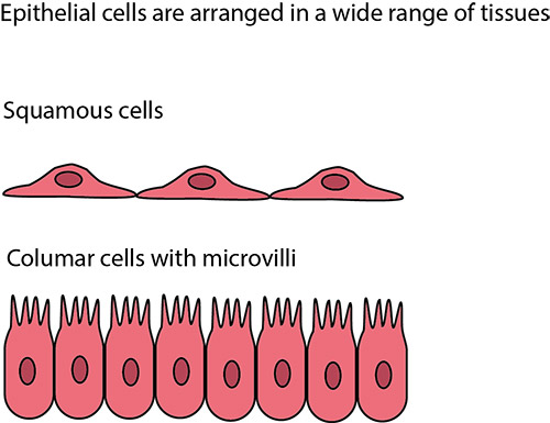
Skin, and the layers within, are important as the largest organ of the body and the wide range of conditions that may afflict it. Examples are skin cancers and immune disorders. Keratinocytes are the primary cells with the epidermis. But epithelial cells also made up the surfaces of various organs with complex 3D arrangements of cells tightly packed in sheets, often with invaginations and other spatial arrangements. These all act to serve as protective barriers to the tissues within, but have many other functions such as secretion, sensitivity to contact, adsorption and transportation. Due to these complexities, accurate epithelial research models are necessary since the spatial organisation and functions of skin are not replicated in cell monolayers.
As a sample gets thicker than a monolayer, or several layers of cells, imaging can become more challenging. Light within the visible range of many common fluorophores gets absorbed and scattered as the depth of tissue increases. Additionally, the effects of autofluorescence within the visible spectrum increase the noise background and collectively act to attenuate the signal. For this reason, working with probes and illumination towards the NIR range, which suffer less of these effects is often desired. Another technique that can be used is tissue clearing in which the tissues are treated so that the diffractive index is kept more consistent throughout the sample improving image resolution and contrast. High QE imaging cameras and confocal systems help researchers image thicker tissue samples into the millimetres range.

© Oxford Instruments 2026
