Resources
 Part of the Oxford Instruments Group
Expand
Collapse
 Part of the Oxford Instruments Group
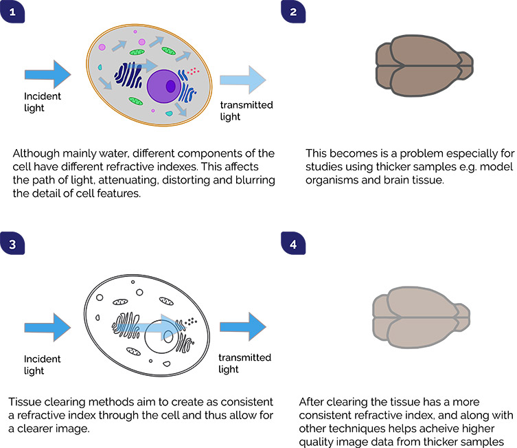
As studies move to larger and deeper 3D samples such as brain sections, organoids and a range of model organisms, it creates a new set of challenges. Issues with imaging thicker samples include light scattering, absorption and autofluoresence which can make it almost impossible to obtain the well resolved images we need to observe features we are interested in. There are some ways we can work around this: using NIR probes that will penetrate thicker samples better with less scatter, using a confocal system like Dragonfly or multi-photon microscopy, and trying image enhancement technology. But the root cause of the challenge is the depth of tissue composed of many biological components like water, proteins and lipids that all act to scatter, blur and attenuate the signal of interest.

One way to help image thicker samples was simply to section the sample into thinner slices, analyse each of them and then reconstruct the images. This may not always be ideal: processing the sample is very laborious and tracing the fine structural details of things like axons is difficult without distortion. Another option we now have that tackles this directly is called tissue clearing. Tissue clearing involves a series of steps to chemically modify the refractive index of the tissue to a level similar to the proteins and therefore allow light to pass through with less refraction. The downsides of this approach include that organic solvents and detergents may damage cell membranes and certain lipid-based fluorophores, and that the clearing process itself takes a long time. A further improvement was achieved by the Chung and Deisseroth groups (2013), with a technique based on the use of acrylamide hydrogels (called CLARITY). A range of tissue clearing kits are now readily available from commercial suppliers that improve the speed of tissue clearing. With these kits most common samples which are well under 1 mm thick will be cleared in a few hours, and even (relatively) very thick samples up to 10 mm within 24 hours.
Tissue Clearing is now widely used technique, especially for neuroscience imaging. sCMOS cameras such as the Zyla and the new back-illuminated Sona series are ideal options as they provide high sensitivity, high dynamic range, fast frame rates and wide field of view.
Further Reading
References
Date: February 2020
Author: Dr Alan Mullan and Dr Claudia Florindo
Category: Application Note
