Resources
 Part of the Oxford Instruments Group
Expand
Collapse
Part of the Oxford Instruments Group
Application Notes
Author: Dr Alan Mullan and Dr Claudia Florindo
Published: 01 Feb 2020 · Last updated: 10 Feb 2021
Conventional light microscopy using fluorophores is limited by the diffraction properties of different wavelengths of light. This means that much of what the cells machinery is doing is lost in an unresolved blur beyond the resolution limit. But with super-resolution techniques such as STED, STORM and PALM this barrier of ~250 nm was broken making resolution down to the nanometre level possible. While the resolution that these techniques could achieve was a significant breakthrough, use of specialised “blinking” fluorophores, high illumination intensities and what would be very high end or specialized equipment restricted their wider adoption. So, it is arguably only within a more recent timeframe that we have truly seen super resolution widely accessible to almost any research group.
Some of the more recent techniques and commercial solutions allow microscopists to access super-resolution much more easily and with less specialised equipment. For example, techniques such as SRRF (and it’s variants such as SRRF-STREAM) are particularly applicable to live cell imaging as they work at low illumination intensities, preserving cell physiology, use standard fluorophores for broad application flexibility, and work across many imaging modalities.
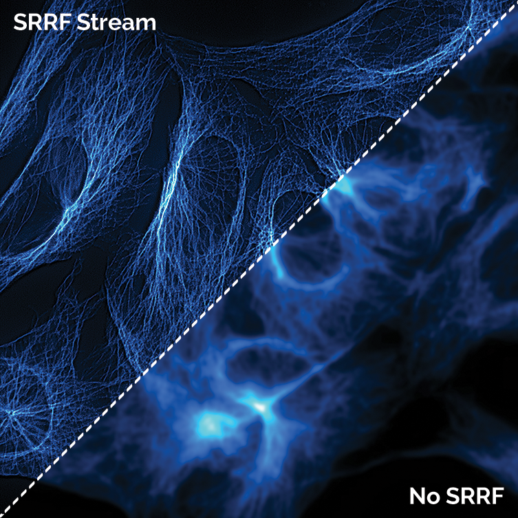
SRRF-Stream readily generates large field of view super-resolution images at fast frame rates. Microtubule structure in fluorescently labelled BPAE cells (Fluocells, ThermoFisher), comparing a widefield image with and without use of SRRF-Stream. The full 1024 x 1024 pixel field of view of the iXon Ultra 888 camera was used. A x63 high-NA objective was used, with further 2x magnification and 560nm illumination. 100 raw ‘input’ images were recorded for every resultant super-resolution image, resulting in a super-resolution image rate of 0.5 Hz. For a fair comparison without SRRF-Stream, 100 standard widefield images were recorded and then averaged .For SRRFStream, a 4x radiality magnification was used, yielding a 4096x4096 pixel super-resolution image.
In terms of super resolution single molecule studies, this is one area where EMCCD cameras still maintain a stronghold. But, sCMOS cameras may also be suitable for many experiments. sCMOS cameras have been used successfully for DNA-Paint and other localisation-based studies. When there are enough photons, the latest back-illuminated sCMOS cameras such as the Sona series are able to provide high speeds and wide field of view so more data can be captured with better temporal resolution.
Further Reading
References

© Oxford Instruments 2026
