Resources
 Part of the Oxford Instruments Group
Expand
Collapse
 Part of the Oxford Instruments Group
In recent work by Hussein et al. in Scientific Reports researchers from 14 institutions collaborated on a research project where Al-Si eutectic solids were imaged using an X-ray projection microscope coupled with a laser-wakefield accelerator (LWFA).1
What is a eutectic system?
A eutectic mixture is a mixture of two substances which solidify homogeneously at a single temperature, which is lower than the melting temperature of either of the constituent components. The physical, mechanical and electrical properties of eutectics are often superior to the properties of the uncombined individual components. Imaging eutectic systems requires high resolution to enable accurate determination of lamella, globular or rod-like structures. In this work an Aluminum and Silicon (Al-Si) eutectic solid are imaged because the lamellar properties of Al-Si eutectic systems were recently resolved using X-ray imaging at the Swiss Light Source (SLS) synchrotron. Thus, the resolving capabilities of the LWFA experiment for microstructures could be easily compared with that of the SLS technique.
What are laser-wakefield accelerators (LWFA)?
Laser-wakefield accelerators provide an alternative route to generating X-ray beams suitable for imaging complex microstructures at high resolution. A high intensity laser is used to illuminate a gas plume creating a plasma wave. The plasma wave results in an acceleration of electrons in a wave that is ~ 1000x more rapid (~100 GV/m) than a typical synchrotron accelerator (~100 MV/m).2 Thus, plasma-accelerated electron beams can produce bright ultrafast bursts of hard X-rays.
Research Overview
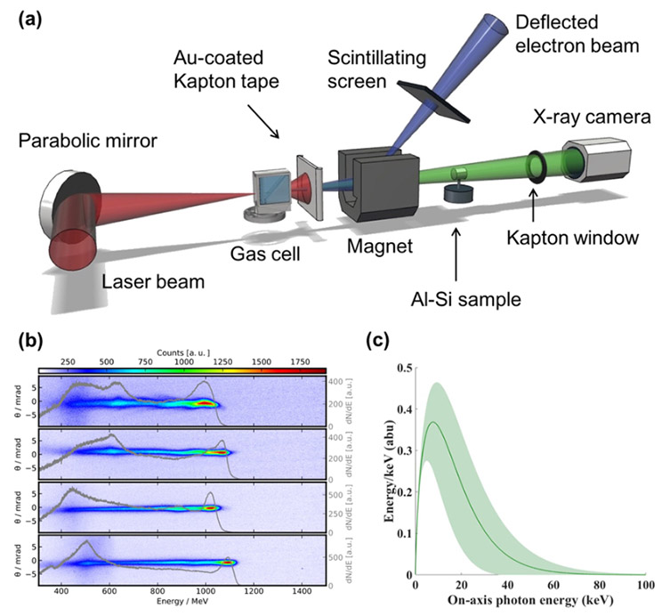
Hussein and coworkers performed the LWFA experiments using the Gemini laser at the Science and Technology Facilities Council (STFC), Rutherford Appleton Laboratory (RAL). An overview of the experimental set up of the LWFA used in this study is shown is Figure 1, imaging was performed using an iKon-L SY detector.

Figure 1: Reproduced from Hussein et al. In a) the experimental schematic is shown with the iKon-L SY as the X-ray detector. A sample of electron beams representative of electron beams used in experiments in this study is shown in b). The betatron X-ray spectrum is shown in c) with an error envelope provided in green.
In Figure 2 a) a microscope image of the machined Al-Si eutectic sample (~ 1 mm) is shown. Additionally, an image obtained using X-rays from the LWFA experiment is presented in Figure 2b); lamellar features at ~ 3 microns are clearly resolvable.

Figure 2. Modified from Hussein et al. In a) Al-Si sample used for LWFA experiments imaged using an optical microscope. Phase contrast X-ray image in b) from LWFA experiment.
Overall Hussein and coworkers demonstrate that high resolution X-ray imaging using the LWFA can resolve lamellar features ~ 3 microns across. Thus, the LWFA could provide an X-ray source which is an affordable and more convenient substitute for synchrotron sources. The X-ray beams generated using the LWFA have low divergence and are less than 100 fs in duration. These ultrafast X-ray beams are excellent for applications which require high spatio-temporal resolution for in-situ measurements. For example, in medical imaging, additive manufacturing and other emerging scientific and engineering applications.
Notes
This work involved researchers from the University of Michigan, Lancaster University, Imperial College London, the Lawrence Livermore National Laboratory, the Gemini laser facility at the Science and Technology Facilities Council (STFC) Rutherford Appleton Laboratory, Diamond Light Source, Lund University, York Plasma Institute, University of Strathclyde, ELI Beamline, Institute of Physics of the ASCR, GoLP/Instituto de Plasmas e Fusão Nuclear Instituto Superior Técnico and the Swiss Light Source, Paul Scherrer Institute.
References
Date: June 2019
Category: Application Note
