Resources
 Part of the Oxford Instruments Group
Expand
Collapse
Part of the Oxford Instruments Group
Application Notes
Author: M. Müller, K. Mann, Laser-Laboratorium Göttingen e.V., Optics / Short Wavelengths, Göttingen, Germany
Published: 01 May 2017 · Last updated: 15 Jul 2025
Tags: tools-tomography
Benefiting from the high absorption contrast between carbon and oxygen, transmission x-ray microscopy in the spectral range of the “water window“ (λ = 2.3 – 4.4 nm) is a powerful tool for the investigation of biological [1] and mineralogical samples [2]. Using Fresnel zone plates as highly magnifying objectives, spatial resolutions in the range of 10 nm have been achieved [3]. However, until now, such studies are almost exclusively conducted at synchrotron sources providing the necessary high photon flux of short wavelength radiation
To enable soft x-ray microscopy for everyday use without waiting times due to limited beam time at large-scale facilities, lab-scale sources based on gas discharge [4] or laser-produced plasmas [5] have been employed. Especially laser-produced plasmas have already been applied successfully in various fields, ranging from material ablation and structuring [6,7] to absorption spectroscopy [8] and transmission x-ray microscopy [9-11]. Here, the most compact soft x-ray microscope is presented operating in the “water window” region with monochromatic radiation from Helike nitrogen at a wavelength of 2.88 nm. It makes use of a nearly debris-free, long-term stable laser-induced plasma source based on a pulsed gas jet.
Experimental
The setup of the table-top soft x-ray microscope is depicted in Figure 1. It consists basically of a laser-induced plasma source, an ellipsoidal condenser mirror, a Fresnel zone plate objective, and a back-illuminated CCD camera sensitive for soft x-ray radiation.
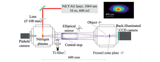
{Figure 1: Schematic drawing of the table-top soft x-ray microscope. The inset shows a pinhole camera image of the laser-induced nitrogen plasma averaged over 10 pulses recorded for the emission wavelength of 2.88 nm}
Due to the high absorption cross-section of soft x-rays in air, the experiments are performed in a vacuum system with a base pressure of 1*10-6 mbar. The laser-induced plasma source has been described in detail elsewhere [12]. It is based on a gas target and a Nd:YAG laser system (Quantel, wavelength 1064 nm, pulse energy 600 mJ, pulse duration 10 ns, repetition rate 5 Hz). The radiation emitted from the nitrogen plasma is filtered by a titanium (Ti) filter (thickness 200 nm) to block out-of-band radiation, such as visible light or scattered laser radiation. Furthermore, radiation below the Ti L-edge (λ = 2.7 nm) is also absorbed, ensuring monochromatic irradiation of samples at λ = 2.88 nm wavelength. The spatial distribution of the plasma is monitored by a pinhole camera (pinhole diameter 50 µm); the nitrogen plasma size at λ = 2.88 nm is measured to be 0.44*0.24 mm² (FWHM).
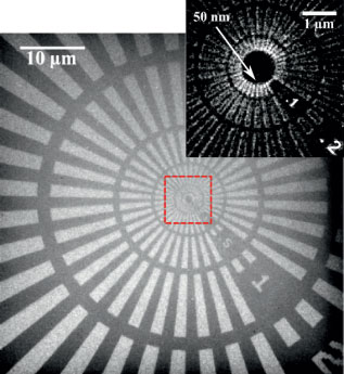
{Figure 2: Soft x-ray micrograph of Siemens star recorded at λ = 2.88 nm (magnification 250x, effective pixel size 52 nm, 18000 pulses, exposure time 60 min). The inset shows the central part of the Siemens star recorded separately at magn. 500x.}
The soft x-ray radiation is collected and focused by an ellipsoidal, axisymmetric nickel coated condenser mirror (Rigaku, Inc., focal length 300 mm, mirror length 100 mm) into the object plane. A Fresnel zone plate objective (ZonePlates Ltd., 190 µm diameter, minimum zone width drn = 30 nm, number of zones 1580) images the sample onto a soft x-ray sensitive CCD camera (Andor iKon-L SO model DO936NOW-BN back-illuminated, 13.5*13.5 µm2 pixel size, 2048*2048 pixels) using magnifications up to 500x
Results and discussion
To assess the imaging performance of the soft x-ray microscope over the full field of view (FOV ≈ 50 µm), initially a Siemens star test pattern was recorded (NTTAT, model ATN/XRESO-50). The corresponding micrograph (see Fig. 2) indicates an almost uniform illumination over the full FOV. As seen from the inset of Figure 2, the smallest structures of the Siemens star having a size of 50 nm are well resolved in all directions.
Further, the polyextremophilic bacterium Deinococcus radiodurans was imaged (see Fig. 3): the representative structure of the bacterium is clearly evident. Other iron enriched micro-organisms were additionally investigated. Due to their iron content they show a stronger absorption compared to carbon containing material. Geo-colloids collected from a water sample (river Main/central Germany) were examined, showing microstructured as well as irregular particles. The necklacelike structure visible in the lower part of the micrograph is supposed to stem from biological debris material. The spacing of the regularly arranged isolated “holes” is approximately 215 nm, indicating the high resolution capacity of the table-top soft x-ray microscope.
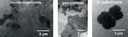
{Figure 3: Soft x-ray micrograph of (left) bacterium Deinococcus radiodurans, (middle/below) iron enriched micro-organisms and (right) geo-colloids taken from the river Main recorded at λ = 2.88 nm (magnification 250×, effective pixel size 52 nm, 18 000 pulses, exposure 60 min). The D. radiodurans was provided by T. Salditt (University of Göttingen), the micro-organisms and geocolloids were collected by J. Niemeyer (University of Göttingen).}
Conclusion and outlook
We have demonstrated the feasibility of a table-top soft x-ray microscope operating at a wavelength of 2.88 nm. It is based on an almost debris-free, longterm stable laser plasma produced in a gaseous target. Currently, structures of about 50 nm in size are already detectable, among other factors limited by the low signal-to-noise ratio due to the relatively low brilliance of the plasma. However, the presented system offers various opportunities for the scalability of the photon flux, which has already been separately investigated in the past. First of all, lasers of higher repetition rate (average power) and / or shorter pulses can be employed. Due to the higher degree of excitation the use of ps laser pulses of the same energy as the currently employed ns pulses can boost the brightness of the plasma by more than a factor of 10 in the “water window” range [13]. Moreover, higher particle densities by use of either higher gas pressures or the barrel shock approach can increase the brilliance of the laser plasma by at least one order of magnitude [14]. Such improvements will be incorporated into the system in the future to reduce exposure times for recording of micrographs, thereby maintaining the compactness of the microscope and especially the inherent cleanliness of the soft x-ray source.
Acknowledgment
Financial support by the “Deutsche Forschungsgemeinschaft” within “Sonderforschungsbereich 755” “Nanoscale Photonic Imaging” (Project C04) is gratefully acknowledged. The authors like to thank T. Salditt and J. Niemeyer (both Univ. Göttingen) for stimulating discussions.
References

© Oxford Instruments 2026
