Resources
 Part of the Oxford Instruments Group
Expand
Collapse
Part of the Oxford Instruments Group
Solution Note
Author: Dr Colin Coates
Published: 01 Nov 2018 · Last updated: 29 Jul 2025
Tags: neuroscience
Challenge Background
Calcium imaging is commonly used across neurobiology to monitor the activity of cells such as neurons or glial cells. Fluorescent indicators such as fura-2, fluo-4 or Genetically Encoded Calcium Indicators (GECI) can be used to directly visualise changes in free Ca2+ levels in response to various physiological conditions or stimuli. When combined with methods such as confocal, 2-photon microscopy, SCAPE or light field microscopy it is possible to look at the localisation and dynamics of neuronal activity from an individual synapse or dendrite, up to the synchronous action of multiple cells as part of the wider neural network. The understanding of these processes can be applied to many areas across neurobiology such as in the development of novel treatments, including those for neurodegenerative disorders such as Parkinson’s or Alzheimer’s.
Cell signalling events are typically highly dynamic, both in terms of the speed at which they happen, and the signal intensities involved. This makes the choice of detector for imaging very important as not only do we have to achieve high temporal resolution, but we also need to have a good spatial resolution, and ability to handle the change in signal intensity level during measurement.
Do you need the widest possible field of view or widest possible dynamic range? Check out the new Sona Back-illuminated sCMOS camera with a 32 mm Field of view and 95% QE.
Technology Solution
sCMOS cameras have the potential to deliver high frame rates and field of view making them perfectly suited to calcium imaging experiments that demand excellent temporal resolution. Previous camera technologies such as CCD and EMCCD are restricted in both speed and spatial resolution. The latest sCMOS cameras offer exceptional sensitivity and the ability to handle the dynamic images in the order of 100s of frames per second (fps) that this work entails.
Andor Camera Solutions for High Speed Calcium Imaging Studies
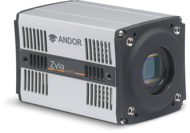
Andor strongly recommend the Zyla 4.2 PLUS with 10-tap Camera Link connection for experiments in which speed is the most critical parameter. Zyla 4.2 PLUS with Camera Link offers the fastest speeds available with 101 fps at full resolution (2048 x2048). Speeds can be boosted even further through use of user definable ROIs and “high speed” mode with up to 1 627 fps at an ROI of 128 x 128. Zyla also provides a wide field of view and high sensitivity from its 4.2 Megapixel sensor with 82% QE.
| Key Requirement | High Speed Calcium Imaging Solution: Zyla 4.2 Plus with Camera Link |
| Achieve excellent temporal resolution of cell signalling events. | Zyla 4.2 PLUS with Camera Link is the camera of choice when your experiments call for the fastest frame rates - along with good field of view and sensitivity. At full resolution the Zyla 4.2 PLUS with Camera Link runs at 101 fps. By using ROIs, speed can be boosted even further: 406 fps at 512x512 and 1627 fps at 128 x 128. This is 3-5 times faster than the fastest EMCCD camera. Result – Enhanced temporal resolution of cell signalling events. |
| Image structures such as synapses, axons, and dendrites | Zyla has a 4.2 Megapixel format sensor with 6.5 micron pixels and high sensitivity which can provide excellent spatial resolution at 60 x and 40x magnification. Result – Achieve superb spatial resolution and localisation within cells. |
| Observe and quantify large changes in signal level. | A high dynamic range of 33 000: 1 and better than 99.8% Linearity means that you can accurately quantify and compare even highly dynamic images. Result – Have confidence in your experimental data. |
| Record accurate physiology of in vivo studies using GECI and other markers. | Zyla has exceptional sensitivity due to 82% QE and a very low read noise of only 0.9e- .This lets you reduce exposure times and fluorophore concentrations for less photobleaching and phototoxicity, preserving accurate physiology of the underlying cell signalling processes. Result – Obtain accurate physiological data for in vivo studies. |

© Oxford Instruments 2026
