Resources
 Part of the Oxford Instruments Group
Expand
Collapse
Part of the Oxford Instruments Group
Application Notes
Author: Dr Marcin Barszczewski
Published: 01 Nov 2018 · Last updated: 05 Aug 2025
Tags: ikon-ccd-long-exposure, In Vivo Imaging
Challenge Background
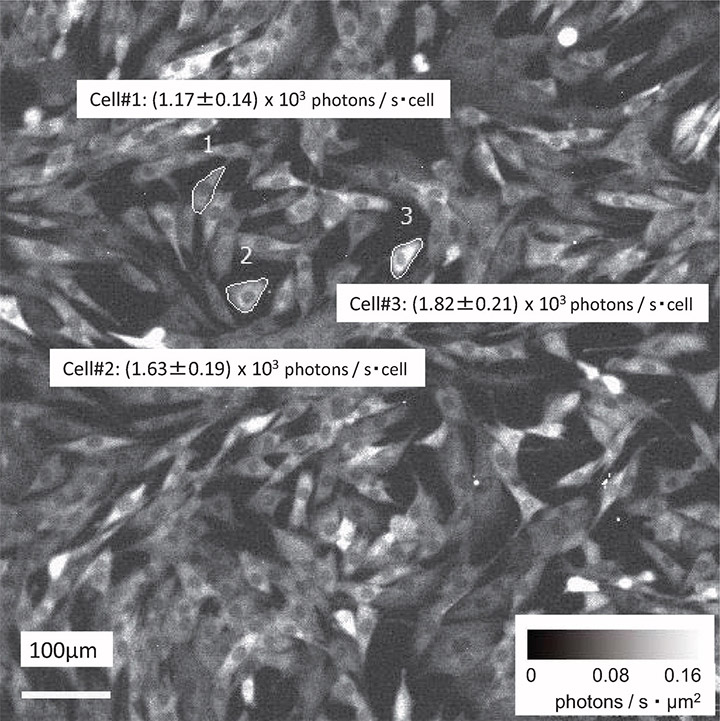
Bioluminescence imaging involving luciferase has been widely used as a reporter system for a wide range of intracellular mechanism involving gene expression and organelle dynamics in single-cell experiments. Such processes may not be suited to fluorescence imaging using one of many types of fluorescent proteins or direct/indirect immunocytochemistry with fluorescently-labelled antibodies. This may be related to complexities involving fusion gene products, steric hindrances related to antibodies or similar challenges leaving bioluminescence as the tool of choice. Bioluminescence imaging presents a distinct set of challenges when done with a CCD-based system. Not only light fluxes in these scenarios are extremely weak - down to single photons per exposure, but the values they produce are relative between measurements sometimes involving different emission wavelengths thus rendering comparisons between separate datasets difficult. Using reference datum of standardised LED emitter together with a highly-sensitive iXon EMCCD camera creates a system that can be a calibration tool for bioluminescence data acquired in different conditions for downstream, like-for-like data comparisons.
Technology Solution
Electron Multiplying CCD (EMCCD) cameras with their precisely controlled thermoelectric cooling and EM gain make them the ideal detector solutions for bioluminescent imaging where photon fluxes from luciferase may be as low as 0.05 photons per µm2 of the sensor. The quantitative stability of EM gain across the entire range of camera readout speeds, pre-amplifiers and temperature settings is a must to ensure reliable quantitative performance throughout a bioluminescent kinetic acquisition and repeatability between measurements.
Andor Camera Solution for live-cell super-resolution
Andor strongly recommends the iXon Ultra or iXon Life Back-illuminated EMCCD Cameras for single-cell bioluminescence experiments. The iXon Ultra and Life are well-established EMCCD platforms with their linear RealGain™ technology that enables unparalleled stability needed for light-starved, high-sensitivity imaging. EM gain’s stability, augmented by iXons’ thermostatic precision of +/- 0.01°C enable users to perform the most challenging bioluminescent experiments
| Key Requirement | Low-light bioluminescence imaging: iXon Ultra 897/888 |
| Sensitivity across broadband visible bioluminescent emission spectrum | Single photon sensitivity combined with EM gain, > 90% QE and tens to hundreds of frames per second (with ROI or Crop Mode) mean that user can capture extremely low emissions from luciferase-derived reporter systems. Result – detection of weakest signals that can be used either as direct measurements of as a part of a tool for establishing absolute intensity reference. |
| Quantitative stability of EM gain | Detailed, multi-facetted optimisation of EM gain control as well as other key camera performance parameters enables the collection of high-quality data for analyses of bioluminescent signals. Result – image data you can trust regardless of the wavelength of bioluminescent emission, EM gain, acquisition speed and other crucial experimental settings. |
| Flexibility of use | iXon EMCCD camera can be optimised for speed and sensitivity depending on your experiments - a perfect fit complementing high-end optical microscope and lenses. Result – a powerful imaging tool suitable for subtly different bioluminescent experimental setups. |
| Simplicity of technical solution | iXon EMCCD camera is compatible with most modern microscopes and their software controls - with its C-mount and appropriate software drivers, it can be deployed on your existing microscope. Result – perform quantitative bioluminescence measurements on your familiar microscope with minimal installation downtime |

© Oxford Instruments 2026
