Resources
 Part of the Oxford Instruments Group
Expand
Collapse
 Part of the Oxford Instruments Group
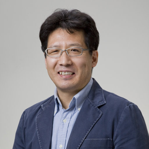
Dr Kazuaki Yoshioka is a staff researcher at Department of Physiology (Prof. Yoh Takuwa’s Lab.), Kanazawa University Graduate School of Medical Science, in Japan.

Dr Kazuaki Yoshioka did his PhD at Tokyo University of Agriculture and Technology in the lab of Prof. Hideaki Matsuoka. Further, he undertook a postdoctoral researcher role at Bioanalytical Systems Inc. (BASi) in the USA; he then moved for a second postdoctoral researcher position at Tokyo Metropolitan Institute of Medical Science. Currently, Dr Yoshioka research focus is in the field of vesicle trafficking.
Dr Yoshioka aims to understand the roles of PI3 kinases in vesicle trafficking, as well as their importance in normal homeostasis and disease. Dr Yoshioka, has an outstanding publication track record, with an H-index of 27 given from 57 publications in the web of science that were cited 1862 times.
Following Dr Yoshioka interesting work on vesicle trafficking, we wanted to understand what motivates his vesicle trafficking research and what are the challenges that he needs to overcome to be able to answer his research questions. Here, we present an interview on Dr. Yoshioka’s recent challenges and discoveries.
Q1: Why are you interested in researching vesicles?
We have been investigating the physiological basis of class II phosphoinositide 3-kinase (PI3K-C2α) in vascular biology. In 2012, we published a paper in Nature Medicine that we found that PI3K-C2α is required for the endocytosis of certain membrane proteins, such as VEGF (vascular endothelial growth factor), S1P (sphingosine-1-phosphate), TGFβ (transforming growth factor-β) receptors, to regulate vascular barrier function. For this reason, we are very much interested in the molecular and cellular mechanisms of endocytic vesicle formation regulated by PI3K-C2α.
Q2. What are the main challenges in vesicle imaging?
I want to try to observe the single endocytic event with multicolour (3-4 colours), high-speed (< 500ms - 1-sec interval) and at high-resolution in a living cell.
I know that TIRF microscopy is one of the best choices to see the dynamics of endocytosis at the cell surface. But we need to follow them until lysosomes via early/late endosomes. Unfortunately, TIRF can image only the initial event of endocytosis in a thin subsurface layer.
Q3. What resolution is required to image vesicles and, to answer your research questions?
We need high-resolution to see clathrin-coated vesicles (CCVs). Formation of CCVs consists of many players such as dynamin, AP-2, actin fibres, phosphoinositides etc. To discriminate those molecules during endocytic events, I need to acquire with 50 -100nm resolution for multi-colour 3D live imaging.
Q4. Why was imaging with SRRF important?
Because, due to low photo-toxicity, SRRF coupled with dragonfly (spinning disk)/iXon EMCCD can extend conventional confocal microscopic techniques to image multi-colour channel with z-stacks for a relatively long period.
Q5. What acquisition speed do you need?
We've taken SRRF images (2-color, 3 x z-stack) of live cells around 10 - 15-sec interval.
Q6. Do your samples undergo photobleaching?
Not so much. It is quite better than I expected. I knew the advantage of the Dragonfly because I’ve been a user of Yokogawa CSU + iXon EMCCD system.
Q7 So as you’ve mentioned you see very low levels of photobleaching, do you think this is be-cause you’re using spinning disk system?
Yes. Generally, the spinning-disk confocal system provides not only high-speed imaging but significantly reduced phototoxicity and photobleaching because of reduced laser exposure. The problem would be that captured images are dark. This problem is overcome using highly-sensitive cameras such as iXon EMCCD!
So, I mean Dragonfly (and also Yokogawa CSU coupled with iXon EMCCD) is, in fact, be the best tool to choose for low photo-bleaching live-cell imaging.
Q8 If you compare a spinning disk system to a point scanner, what would be the advantages?
The spinning disk system has mainly two advantages over a conventional point scan system: “High speed” and “lower photo-toxicity and -bleaching”.
Q9 Do the samples bleach very much in a point scanner? And in a widefield system?
Yes, very much indeed. I never use those systems for live-cell imaging.
Q10. Why did you favour using the dragonfly instead of your existing CSU system? What advantages does the dragonfly have that lead you to this choice?
Both Dragonfly and CSU system are good systems for live-cell imaging. But our Yokogawa system (CSU10 + iXonEMCCD DV887) is an old setting. We've suffered from its low resolution and sensitivity. Dragonfly helped us with those issues. The best thing of all, Dragonfly can simply give us Super-resolution images.
Q11. Would a combination of TIRF and SRRF be beneficial in your studies?
Yes, I wish I could. I realise that live-imaging using Dragonfly’s SRRF combined with TIRF channels would be the most powerful system to have an impact on Life science.
Q12. What features of the Dragonfly help you most in your research?
When trying to determine the subcellular localisation of our interest(s) within a cell, Dragonfly allows us to perform multi-colour (DAPI-, GFP-, RFP- and Far-red channels) 3D imaging with continuously high-resolution (confocal)-wide view mode and super-resolution (SRRF)-magnified view mode. From practical experience, I can say it has absolutely incredible features!
Q13. Are there any moments that stand out while producing this data?
I was impressed with Dragonfly’s performance that provided us with an almost perfect balance of resolution and speed. Dragonfly can be applied to a diverse range of biological samples from a single live cell to whole tissue sections.
Q14. Any other notes that you would like to add?
I love the Dragonfly. Why should I go for old microscopy?
Click here to read our application note on Dr Kazuaki Yoshioka research work.
