Resources
 Part of the Oxford Instruments Group
Expand
Collapse
Part of the Oxford Instruments Group
Case Studies
Author: Dr Claudia Florindo
Published: 01 Nov 2019 · Last updated: 19 Dec 2022
Challenge Background
Mitochondria provide cell energy and thus, are essential organelles of the eukaryotic cell. Defects in mitochondria dynamics are associated with acute syndromes such as neurodegenerative disorders, cardiovascular disorders and neurometabolic diseases.
Microscopy in mitochondria is demanding, these organelles have a complicated inner architecture with two membranes, a smooth outer membrane and an extremely tortuous inner membrane. Besides the membrane architecture mitochondria are small, with a size near the resolution limit of the light microscopes. Moreover, mitochondria are extremely dynamic organelles that divide, fuse, change shape and travel along the intracellular space. All these characteristics pose obstacles for imaging live mitochondria for studying their dynamic behaviour (fission and fusion) as well as the underlying rapid signalling processes that lead to fusion or fission.
When imaging live mitochondria, besides the critical challenges of live imaging, including phototoxicity and photobleaching, other problems need to be overcome. Images must be obtained with high temporal and spatial resolution. In fact, to achieve the most comprehensive picture of the spatial and temporal mitochondria dynamics, it is necessary to acquire the image data in 4 dimensions (3D + time). Even for fixed mitochondria, high spatial resolution is an essential requirement due to their size and complexity.
Technology Solution
A camera-based confocal system is a better solution for live mitochondria imaging that does not compromise cell viability due to phototoxicity. The equipment will need to acquire at fast speeds and with high imaging resolution.
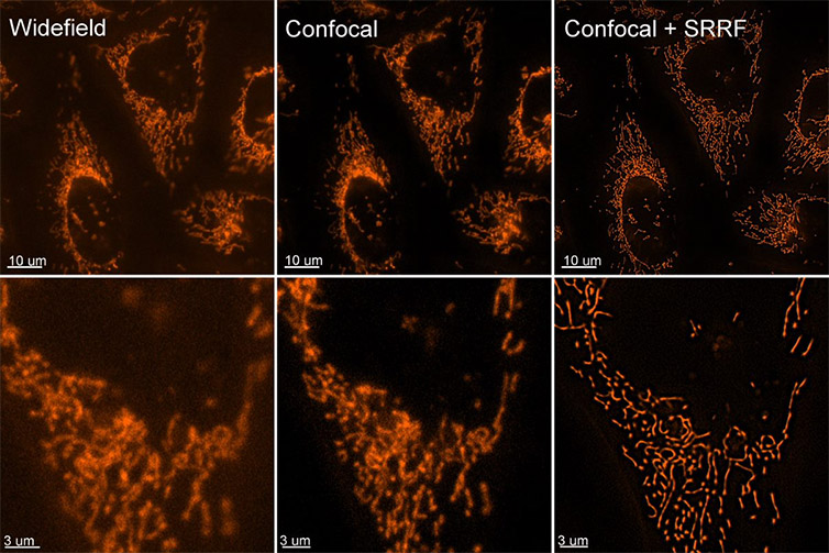
Figure 1 – Images of live cells stained with Mito Tracker acquired in widefield, confocal and confocal + SRRF stream. An increase in resolution can be observed between the widefield, confocal and confocal with SRRF stream images. Using Dragonfly with SRRF-stream, scientists can acquire live-cell super-resolved images of mitochondria.
Andor Solutions for Imaging Mitochondria
Dragonfly is the complete solution to image live mitochondria due to its speed, sensitivity and resolution. Highly sensitive cameras allow the detection of very dim signals with high Quantum Efficiency. Ultra-fast low-light imaging is possible with Dragonfly and Andor´s cameras such as the back-illuminated Sona or iXon EMCCD series. Imaging modalities such as confocal spinning disk, dSTORM super-resolution (resolution ~ 20 nm) are possible with the Dragonfly, and each of these imaging modalities has advantages for mitochondria studies.
Importantly any modality can be combined with the super-resolution technique SRRF (super-resolution radial fluctuations). SRRF-stream is compatible with live-cell imaging, offering the advantage of high-speed super-resolved live imaging.
| Key Requirement | Mitochondria Imaging Solution: Dragonfly and Andor´s high QE cameras |
| Ultra-fast acquisition speeds | The EMCCD and sCMOS detectors in the Dragonfly allow very low light imaging and high acquisition speeds. Dragonfly is at least ten times faster than point scanning confocal systems. The internal beam splitters allow Dragonfly to acquire two channels simultaneously on two independent detectors (cameras). Result 1 - Detect ultrafast events with acquisition speeds up to 400 frames per second. Result 2 - Detect two independent channels simultaneously without compromising speed or resolution. |
| Acquire images with a resolution of 50-100 nm | Dragonfly supports dSTORM. Dragonfly also has a motorised astigmatic lens that creates a calibrated asymmetric distortion of the single-molecule PSF, which varies with axial defocus. Result 1 - Resolution down to 20 nm with dSTORM. Result 2 - Acquire 3D super-resolved images combining dSTORM and astigmatic lens. |
| Acquire live Super-resolved Images (resolution Up to 100 nm) | Andor´s cameras offer an integrated licence for SRRF (Super-resolution radial fluctuations). SRRF uses the fluctuation of fluorophores emissions and their interpolation to increase the effective resolution of an optical system. Andor's built-in SRRF stream algorithm allows real-time SRRF calculations and immediate visualisation of the resolution increase. Furthermore, SRRF is compatible with conventional fluorophores, and there is no need for complex sample preparations. SRRF is compatible with confocal, TIRF and widefield imaging, and with deep imaging. Result 1 - SRRF-stream yields an increase of resolution between 2- and 6-fold (50-150 nm final resolution) in the final data. Result 2 - Due to its low power requirements (mW/cm2 to W/cm2 range), SRRFstream is compatible with live-cell imaging. Result 3 - SRRFstream algorithm allows acquisition of live cell images that break the diffraction limit at a frame rate of 10 frames per second. |

© Oxford Instruments 2026
