Resources
 Part of the Oxford Instruments Group
Expand
Collapse
 Part of the Oxford Instruments Group
Facilities such as synchrotrons and X-ray Free Electron Lasers (XFEL) have been the mainstay of UV and X-ray research for decades. Recent improvements to High Harmonic Generation (HHG) based sources are now offering powerful, affordable laboratory-based tools for probing a wide variety of samples with radiation spanning from UV to soft X-Ray. All three of these methods allow experimental analysis of samples that cannot be achieved with light sources in the visible range and require UV and X-ray light to provide unique insights into the complex structural and/or elemental make-up of, for example, engineered and biological samples with the highest degree of accuracy.
As well as UV and X-ray beamlines, Neutron sources are another tool in high energy physics laboratories. With spallation and nuclear sources, the most common types of neutron producing beamlines. Neutrons are used for imaging low atomic number element samples that high attenuation characteristics through absorption and scatter.
UV and X-ray photons have higher energy and therefore shorter wavelength than visible light. We can further classify UV light into Vacuum UV (VUV) and Extreme UV (EUV, XUV). X-rays can be classified into Soft and Hard X-rays. This higher energy light needs specific detection methods for direct detection and indirect detection. For indirect detection, scintillators are used which convert the high energy photons into visible photons, whereas direct detection is done without the use of a scintillator.
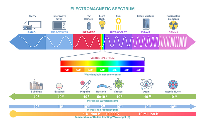
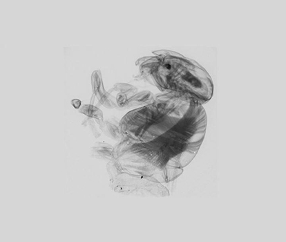
UV and X-ray light allows scientists to probe deeper into materials than is possible with visible wavelengths. This can be seen with medical X-rays where different material densities are visible on an X-ray radiograph due to their different attenuations of the X-rays. The same properties can be used by scientists when imaging biological or material samples. Modern experiments often involve single radiograph imaging or can combine multiple images to form a 3D tomographic image.
High energy light is also used in diffraction and scattering experiments where the structure of samples is probed down to the atomic level. These experiments are often run at dedicated beamlines at synchrotrons, XFELs or with HHG sources.
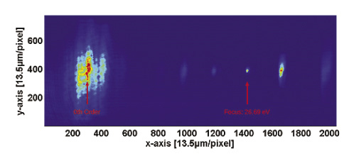
Many beamlines have recently been upgraded, are currently going through upgrades, from so-called Generation 3 to Generation 4 types, or have upgrades planned. These upgrades increase the repetition rates of the sources and increase the brilliance of the light they emit. New camera technologies are now required that work at higher frame rates, to match those of the sources, whilst maintaining accuracy and a low noise floor without sacrificing dynamic range to provide data with the highest contrast and accuracy.
Neutrons have different interaction mechanisms with matter than X-rays so offer a complimentary imaging technique. As with X-rays, neutrons can also be used in Computed Tomography (CT) experiments to reveal the 3D structure of samples. Neutron-based diagnostics can provide information on the inner structure, damages, authenticity, manufacturing and restoration processes of cultural heritage artefacts including for example metal objects (e.g. lead, bronze or iron-based weaponry or statues), ceramics, or samples combining metallic and organic materials (e.g. wood, adhesives, soil, leather, lacquer). Other techniques such as Bragg-edge, Small Angle Neutron Scattering (SANS) or Grating Interferometry imaging techniques are also typically used to identify strain/stress/cracks in bulk materials for sample diagnostics.
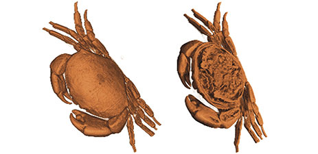
Many neutron sources are also in the process of being upgraded with new beamlines and higher energy neutrons being produced more often. This is driving new camera technologies that are able to work at higher frame rates whilst maintaining accuracy and a low noise floor without sacrificing dynamic range to provide data with the highest contrast and accuracy.
The initial driving laser beam that an HHG beamline uses can be one of a variety of wavelengths. With the wavelength of these driving lasers typically ranges from 800nm to 2100nm [1][2][3]. Beam profiling can be carried out on this initial driving laser as a diagnostic tool [4] as well as used for infrared imaging at this point of the beamline [4]. By providing diagnostic checks on the seeding light source and the light source itself, the system can be optimised to produce the best quality UV and Soft X-ray beam as an output.
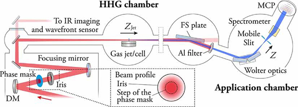
The best camera to suit the HHG infrared diagnostic and imaging applications depends on the exact wavelength of the driving laser beam. The C-BLUE ONE is optimised for the more visible end of the spectrum at 400 to 1000nm and features a peak QE of over 70%. As we move deeper into the Infrared range we move to the C-RED range of cameras where the C-RED 2, C-RED 2 Lite and C-RED 3 all offer a peak QE of over 70% and work in the wavelength range from 0.9 to 1.7 µm. The C-RED 2 also comes with extended range options for even longer wavelengths. The C-RED 2 ER has two options available, one for the wavelength range 1.1 to 1.9 µm and 1.3 to 2.2 µm.
The C-BLUE ONE comes with a range of sensor options which includes 0.5 MP, 1.7 MP and 7.1 MP sensors. The 0.5 MP version of the C-BLUE ONE offers maximum speed with up to1594 fps for full sensor imaging and features 9 µm pixel pitch.
Whereas the 7.1 MP version offers smaller pixel pitch at 4.5 µm but a larger sensor diagonal at 17.6 mm compared to the 9.2 mm of the 0.5 MP version. This 7.1 MP sensor comes with an impressive maximum readout speed of 207 fps.
The C-RED range of cameras have a 640 x 512 pixels array with a 15 µm pixel pitch and up to 600 fps across the standard and Extend Range detectors.
First Light Imaging cameras offer driving laser beam profile diagnostics with both the C-BLUE ONE and C-RED 2 range of cameras. This product portfolio covers a wide wavelength range from 400 to 2200 nm and features a variety of pixel pitches and array sizes to match the needs of the experiments.
Experimental beamlines are tuned to different wavelengths to probe matter in different ways. Many UV beamlines are used to perform spectroscopy [5][6][7] on a wide variety of samples to determine, for example, their elemental composition and behaviour in different environments. These beamlines need a detector that matches the centre wavelength of the UV light created by the source. Direct detection technology is often used for UV light with many silicone-based detectors working in this wavelength range.
The C-BLUE One UV has additional QE in the 200 to 400nm region than that of the standard C-BLUE One camera. This means it is ideal for beamlines that work in this wavelength region. The 8.13 MP sensor comes with 2.74 µm pixel pitch offering a high resolution sCMOS sensor whilst offering frame rates of up to 170 fps to gather maximum data from the experiments.
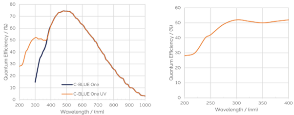
The C-BLUE One UV offers direct detection UV imaging down to 200 nm whilst offering a high resolution 8.13 MP sensor array with 2.74 µm pixel pitch. This high accuracy sensor comes with a maximum full frame imaging speed of 170 fps to offer a high-speed, high-accuracy detector for UV imaging.
As we move into higher energies of UV light and into the X-ray region or neutron detection, it is necessary to move to sCMOS detectors with indirect detection and the additional use of scintillators. These scintillators convert the incident high energy photons or neutrons into visible photons that can be detected by the sCMOS camera. The use of scintillators allows detection of X-ray energies up to 100 keV and neutrons up to the MeV range, well beyond the detection limit of direct detection camera technology.
With many beamline facilities in the process of being upgraded or recently completing upgrades, neutron and X-ray sources are now running at faster repetition rates and higher brilliance than ever before. This means that the sCMOS detection needs to run at higher frame rates to match these sources. The C-BLUE One offers full frame imaging up to 1594 fps and has a QE of over 70% with a curve that matches many available scintillators to maximise the detection of these visible photons.
The C-BLUE One is available with sensor arrays up to 7.1 MP and pixel sizes down to 4.5 µm. This allows the optimum sensor to be matched to the resolution and field of view of the beamline experiment.
The C-BLUE One range offers an extremely high-speed indirect detection sCMOS sensors to be used at modern beamline facilities. The flexibility of sensor choice allows scientists to match their detection requirements to their specific experimental needs in terms of resolution, field of view and full frame imaging speeds whilst having the additional benefit of high QE across a range of frequencies, allowing flexibility in the choice of scintillator used for indirect detection.
Take home message
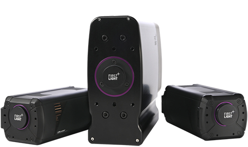
First Light Imaging cameras offer detection solutions that meet the needs of a wide range of beamline facility requirements. From infrared detection (C-RED range) for HHG beamline monitoring, visible detection (C-BLUE ONE) for imaging neutron and X-ray scintillators to direct UV detection (C-BLUE One UV) down to 200 nm. To see the full range of First Light Imaging cameras and Oxford Instruments Andor cameras for high energy physics laboratory based and large facility beamlines, click here.
First Light Imaging cameras are designed to cover a wide range of the spectrum whilst offering low noise and detector flexibility to match experimental needs. Data communication interfaces have been chosen to optimise throughput whilst minimising induced noise from the tough detector environments found at beamline facilities and to offer data transmission over large distances.
For X-ray and neutron imaging at high frame rates, sCMOS detectors work with scintillators in beamline experiments. The C-BLUE One was developed with a range of high-accuracy sensor options to meet these experimental needs whilst offering high frame rates to meet the speed requirements of ever-increasing repetition rates of sources. Scintillators such as EJ254 are optimised for a broad neutron energy range and emit blue light (425 nm) whereas more tradition scintillators emit more in green (550 nm). The C-Blue One has high QE across this range allowing it to be used with a wide range of scintillators.
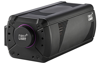
For lower energy beamlines in the UV region of the spectrum, the C-BLUE One UV has been developed to offer direct detection down to 200 nm. The 8.13 MP pixel array combined with 2.74 µm pixel pitch, creates a high-accuracy sensor capable of high-speed full sensor imaging up to 170 fps.
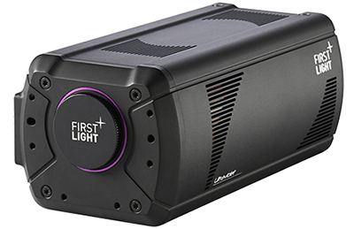
Thanks to their high performance in the infrared spectrum, the C-RED range of cameras are a great tool for driving laser monitoring and infrared imaging. The C-RED 2 Extended Range (ER) option further expands the wavelength range to match the longest wavelength driving lasers commonly found in HHG experiments

Beamline facilities have a range of imaging requirements that cover a broad range of the spectrum. This combines with the requirement for high-speed detection, to match current and next generation beamline sources, whilst maintaining high resolution and large field of view.
First Light Imaging develops a range of cameras to meet beamline detection requirements. In the visible range, the sCMOS C-BLUE One can be combined with scintillators for X-ray detection up to 100 keV. As we move to UV wavelengths, the C-Blue One UV offers direct detection down to 200 nm which is particularly useful for HHG experimental setups. The C-RED range covers the infrared spectrum which makes it ideally suited for HHG driving laser monitoring and imaging in these longer wavelengths.
Date: October 2024
Author: Dr Joe Brennan
Category: Application Note
