Resources
 Part of the Oxford Instruments Group
Expand
Collapse
Part of the Oxford Instruments Group
Technical Article
Author: Dr Alan Mullan & Dr Aleksandra Marsh
Published: 01 May 2020 · Last updated: 10 Feb 2021
HeLa cells are the most well-known and widely used in the biological research community. The name HeLa comes from the patient Henrietta Lacks; whose cancer cells were isolated by Dr George Gey in 1951 to become the first recognized immortal cell line.
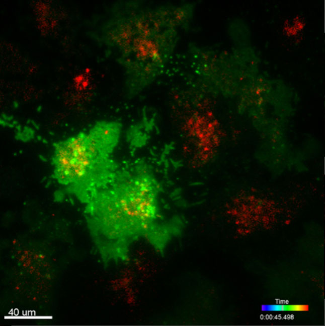
Hela cells expressing GFP fusion protein and labelled with DiI, which is retained in the lipid bilayers of the cell membrane. The TIRF technique provides and enhancement in contrast over conventional fluorescence microscopy, but only within a shallow depth.
HeLa cells are the most well-known and widely used in the biological research community. The name HeLa comes from the patient Henrietta Lacks; whose cancer cells were isolated by Dr George Gey in 1951 to become the first recognized immortal cell line.
HeLa cells have played a key role in many of the scientific developments of the last 60+ years including the development of the Polio vaccine, as well as work on HIV and numerous cancer studies.
The HeLa cell line has endured as a research model for the last ~70 years because it can be easily grown, is incredibly robust and is available as a free resource from John Hopkins. Moreover, with the volume of work done on these cells it means that it is well characterised making it possible to infer more information. One issue with HeLa cells is that due to their robust nature they can inadvertently end up as contaminants of other cell lines.
Read more about the origin of HeLa cells and their importance in the biological research community in the book The Immortal Life of Henrietta Lacks.

© Oxford Instruments 2026
