Part of the Oxford Instruments Group
Expand

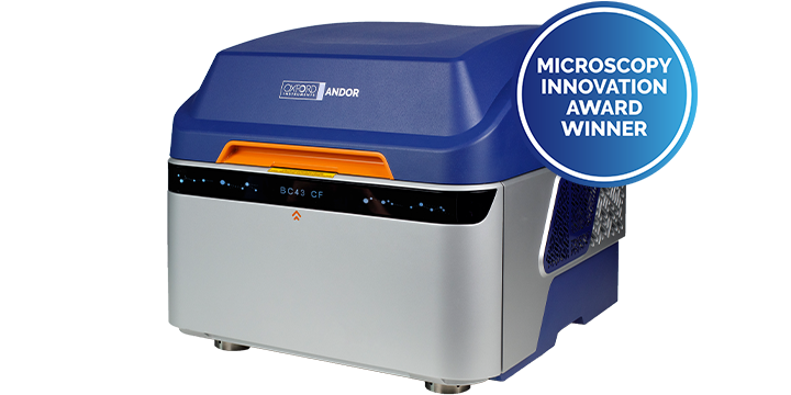
BC43 Widefield and Confocal Benchtop Microscopes

Exceptional performance, Certified quality, High productivity

BC43 benchtop microscopes offer high-quality images, 2D/3D, widefield or confocal. The super-resolution option delivers resolutions down to 140 nm with the push of a button. Its certified quality ensures reproducibility and reliability of data between experiments and different research groups.

BC43 includes Fusion Benchtop software, for straightforward multimodal imaging acquisition, and Imaris, the market leader in image analysis software.

Small in size, Big in performance. 

Download Brochure Request Pricing

Fast, Compact, Upgradable, Premium Microscopy

With certified quality, BC43 offers productivity and image excellence while maintaining the ease of use of a conventional entry-level widefield system, delivering high-end features at a competitive price.

Click the hotspots to see BC43 features and benefits explained.

1
Advanced Imaging Technology
2
Enhanced visualisation software
3
Easy to use
4
Light-tight lid
5
Optimal performance
6
Flexible
7
New Super-resolution Mode
8
Analysis Workflows

Compact - fits on a bench, no need for a dark room, built-in antivibration mechanism.

Super-fast learning curve - new users can be independent within 30 min to 2 h – minimal training and user support needed.

Modular and upgradable - Widefield or Confocal, in-field upgrade , Modular software to adapt to user needs.

Unique & certified quality - IQ/OQ quality control - Certified by experts in the scientific imaging field QUAREP-LiMi*.

Target productivity - dual microlens spinning disk integrated with a sensitive sCMOS camera and specialised SW modules.

Live cell imaging - dual microlens spinning disk, efficient light collection results in gentle imaging, (image model organisms for days).

Versatility - with 4 lasers, 4 imaging modalities and a range of objectives, all your important microscopy lab experiments can be done hassle-free.

Image Analysis – Includes Imaris the gold standard software that delivers image analysis with precision, speed and user-friendly workflows.

Book Demo

* Consortium for Quality Assessment and Reproducibility for Instruments and Images in Light Microscopy

Confocal, Widefield & Transmitted Light Microscopy All-In-One

The BC43 benchtop microscope is offered in 2 models: BC43 Widefield (BC43 WF) and BC43 confocal (BC43 CF). Both models include transmitted light options, DPC and brightfield, and can be upgraded to super resolution. ​

The BC43 widefield is:

The BC43 confocal delivers:

Confocal Microscopy

Speed, Productivity, Image excellence and Quality

 With BC43 confocal, the researchers can:

  • Capture superb confocal images at least 10 times faster than point scanner-based systems
  • Image deep into thick samples in 3D easily.
  • Image live samples for days without phototoxicity or photobleaching.
  • Image fast live events due to a high-sensitivity, high-speed imaging camera.
  • Take advantage of the motorised XYZ stage to increase productivity, either by acquiring multiple points or large organism montages, all in one experiment
Request Pricing

Widefield Fluorescence Microscopy

Image excellence, Certified quality, Field upgradable

 BC43 widefield system:

  • Ideal for imaging light-sensitive and thin samples
  • Offers the fastest imaging speeds to capture dynamic processes in live cells.
  • Detects lower sample signals than in confocal mode.
  • Boosts productivity by the use of: 1) extremely precise XYZ stage, and 2) the power of fusion software.
  • With Clearview™ GPU deconvolution removes sample blur and enhances resolution.

If user a needs grow and a powerful confocal system is required to image deeper, and with increased resolution, the BC43 widefield can be field upgraded to the BC43 confocal.

Request Pricing


After
Before

Transmitted Light Microscopy

Label-free imaging, Enhanced contrast, no Phototoxicity ​ 

BC43 delivers two different transmitted light options for excellent imaging flexibility:

Brightfield Mode to visualise the development of larger organisms, whose structures naturally deliver high-contrast images.
Differential Phase Contrast (DPC) is ideal for transparent samples, allowing the visualisation of structural detail.

Advantages of BC43´s transmitted light methods: ​

  • No need for sample labelling
  • Low energy illumination: no phototoxicity, no photobleaching
  • Reveal full organism boundaries – visualise the spatial context. ​
  • Overlay with widefield or confocal: Integrate protein localisation into the organism's environment
Request Pricing


After
Before

Super Resolution Microscopy

Fast, One click results

We have developed and optimised our popular SRRF-Stream+ technology for exceptional ease of use. Visualise cell structure, protein interactions, membrane fusion, signal transduction events and more.​

SRRF-Stream+ is compatible with:​

  • Standard labelling protocols.​
  • Thick and thin samples. ​
  • Widefield and confocal imaging modalities.​
  • Live imaging protocols. ​

Overcome the diffraction limit by a factor of 1.5x to 1.7 x down to ~140nm.

Request Pricing


SRRF Stream+
Confocal

IQ/OQ Installation Qualification / Operational Qualification

Precision You Can See. Quality You Can Trust

IQ/OQ is a unique and innovative service offering from Oxford Instruments that ensures the performance of all our benchtop microscope systems. Guaranteed, measured performance enables the comparison of experimental results across time and laboratories conducting the same research. Trust your data, validate your results.

  • Certified quality assurance validated by published protocols.
  • Quality certified with protocols developed by Experts in the Field QUAREP -LiMi.
  • Standardised procedures.
“IQ OQ initiative holds great significance for scientific experiments in our field. It promotes standardised procedures, reduces variability, and enhances the reproducibility of results. Ultimately, it contributes to the credibility of research done with microscopes and advances the pursuit of scientific knowledge “

Dr. Glyn Nelson, Core Facility manager – Newcastle University, UK, QUAREP-LiMi Member

Learn More Request Pricing
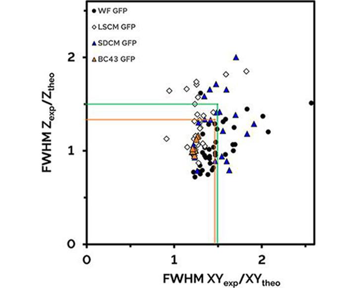
Lateral and axial resolutions of different types of fluorescent microscopes as a ratio of the theoretical full width half-maximum (FWHM).
Acceptable normalized resolution, within green square. Orange square shows that all measured BC43 (BC43 GFP) results are significantly better that the proposed spec of QUAREP-LIMi (green square). Note the variation observed on competing widefield (WF-GFP), laser scanning ( LSCM GFP) and spinning disk (SDCM GFP) competitors products. Figure adapted from data and specifications included in Faklariset al.

High-Speed Confocal Imaging for Biologists

The BC43 Benchtop Confocal has been developed in collaboration with leading biologists; for use across life science applications, and is designed to be part of a single room workflow. BC43 will bring instant clarity and depth to your research, whatever your sample, in ways previously only found in larger, more complex and expensive imaging systems.

BC43 addresses the broad scales and types of imaging protocols and analysis related to cancer research. From subcellular and cellular live cell experiments to large tissue samples and organoids requiring deeper imaging.

  • Image monolayer and 3D cell cultures.
  • Follow cell dynamics (intercellular behaviour, migration and division).
  • High productivity imaging of tissue preparations.
  • Analyse movement, cell lineage, count cells, features and more.
  • Boost your unit with the Super-resolution module for detailed spatial understanding of actin filaments, microtubule networks, distribution of vesicles and lysosomes etc.

Pancreatic ductal adenocarcinoma cells inside and on top of a 3D hydrogel. DNA is blue, lamin A/C is green, and tubulin is orange. Image credit: Sebastian Amos and Yu Suk Choi, University of Western Australia.

Request Pricing

BC43 conquers the diverse range of protocols that Cell Biology experiments demand: from a simple 2D snapshot of a cell to understanding dynamic processes with time-lapse.

BC43's unrivalled clarity and speed will save you time without compromising resolution (X,Y or Z) or field of view.

  • Confocal mode for fast, high-resolution imaging of tissues.
  • Widefield mode for higher sensitivity and maximum speeds.
  • Built-in GPU-based ClearView™ deconvolution technology boosts image clarity.
  • Boost your unit with the Super-resolution module for detailed spatial understanding of actin filaments, microtubule networks, distribution of vesicles and lysosomes etc.

Andor’s Benchtop Confocal is a compact, but fully featured system for routine imaging. It can be the imaging workhorse you are looking for, freeing up your sophisticated systems with advanced tools for those who need them.

Simplicity of use has been designed into BC43 to reduce training time and ongoing support of users, enabling labs and facilities to become more efficient.

Researchers will rapidly become competent in acquiring excellent 3D images, and higher multi-dimensional experiments on their own.

  • Four excitation lines (405, 488, 561 & 640nm).
  • Up to 5 imaging objectives (exchangeable).
  • Multiple imaging modes offers flexibility whatever the sample and whatever your budget and needs.

Cut through the challenges of your 3D world and capture images in perfect clarity - from the very first rounds of cell division to the fully developed organism.

Extract crisp 2D images or explore stunning 3D volumes in a fraction of the time you’re used to. Andor’s BC43 Benchtop Confocal delivers fast, high-resolution imaging of developing model organisms such as zebrafish and Drosophila.

  • Image deeper than conventional fluorescence microscopes.
  • Boost productivity 10-fold compared with traditional confocals.
  • No sacrifices to resolution, field of view or 3D detail for speed.

Break through the burden of countless images demanded by your neuroscience research with a ten-fold speed boost, compared to conventional confocal microscopes. Flexible imaging modes cover sensitive live imaging of neuronal processes to large scale imaging of entire neuronal networks in 3D.

  • Outstanding real-time 3D visualisation for immediate insight.
  • Discover more imaging live with high temporal resolution.
  • Dedicated image analysis tools with Imaris: automated process tracing, auto depth, spine detection and more.
  • Add super-resolution for fine detailed imaging of neuronal processes.

Capture stunning 2D and 3D images from live tissue models to whole cleared-tissue preparations.​

BC43 is a true workhorse instrument for large sample imaging, capturing in a tenth of the time of conventional confocal systems and imaging deeper than widefield epifluorescence with deconvolution or digital clearing.​

  • Time-saving, large-view, high-resolution imaging of tissue.
  • Capture huge samples with tiled volume imaging and stitching​.
  • Innovative analysis tools available with Imaris: machine learning object classification, spatial analysis, volume and morphology and many more.

Fusion Benchtop - Powerful and Intuitive Acquisition Software

User-Friendly. Easy to Learn. Built to Deliver.

At the heart of BC43 is a software that is easy to use yet feature rich, flexible and modular, allowing the system to grow with your research needs. Fusion Benchtop includes features that enhance productivity and provide high-quality image visualisation.

Some of the fusion bench top features include:
  • Multi-dimensional imaging protocols – 3D time lapse or multi-position montage
  • Real-time 3D images – visualise data in 3D while (using the Imaris rendering engine)
  • Deconvolution – activated on the protocol – no need for post-processing
  • Stitching - activated on the protocol - Stitch Montage acquisitions in XYZ
  • Spiral montage – effortless sample overview
  • Irregular montage - acquire only the relevant montage tiles
  • Focus Map – correct for tilt and sample wobbling
  • Z intensity correction - visualise all the details deep inside your sample
Request Pricing

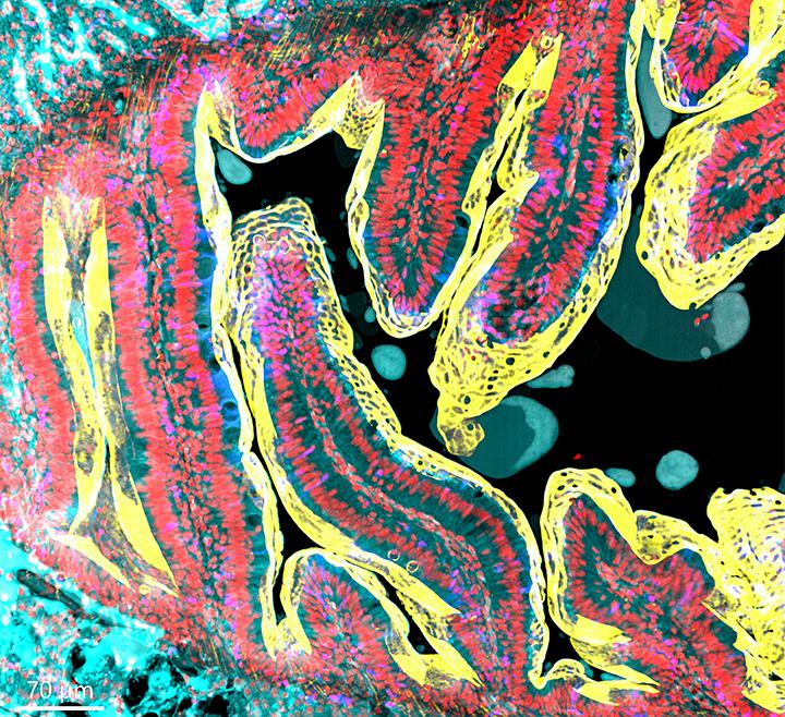
Zebrafish intestine stitched image.
Image was acquired using the confocal imaging modality of BC43, with 4 imaging channels, 77 stacks and 28 tiles. The full stitched image is composed of a total of 8624 images. The deconvolution and stitching options were both activated on the protocol. (image acquired ~ 30 min).
Sample courtesy of Julien Resseguier, at NorMic, University of Oslo. Image credit: Claudia Florindo, Oxford Instruments

Imaris – Market leader in Image Analysis

Comprehensive Image Analysis Software with AI Tools

All Andor Benchtop Microscopes come with the Imaris Quant package for users to:​

  • Visualise their 2D/3D/4D images in the world’s leading interactive microscopy image analysis software. ​
  • Identify and characterize objects within their images via traditional intensity-based and AI methods. ​
  • Report counts, morphology metrics, intensity values, and many more statistics to find differences across images and experiments. ​
  • Create high-resolution snapshots and multi-dimensional movies with ease. ​

Additional Imaris modules are also available including tools for tracking and reporting motility metrics, quantifying cells and their intracellular organelles, and tracing neurons for reporting their specific characteristics.

Learn More Request Pricing

Hear From Our Customers

We image high numbers of thick and large tissue samples demanding a reliable confocal microscope to handle big experiments. BC43 is a true workhorse, requiring minimal experience to obtain high quality images. Being able to put it on a regular bench in our own lab is an efficiency bonus!

Professor Jacco van Rheenen, The Netherlands Cancer Institute.

Professor Jacco van Rheenen
Joaquim Miguel Nunes Vieira

Our research is focused on the cellular and molecular interactions taking place during normal heart development. To gain insight into these we rely heavily on imaging approaches and the BC43 confocal system has been instrumental to advance our studies. Its low footprint combined with its multi-imaging modalities and plug-and-play features are a perfect match!

Joaquim Miguel Nunes Vieira, PhD, King's College London.

Customer Publications