Resources
 Part of the Oxford Instruments Group
Expand
Collapse
 Part of the Oxford Instruments Group
Super resolution microscopy has helped provide greater insights into the fundamentals of cell biology by revealing structural information and interactions that were previously hidden beneath the diffraction limit. Fluorescence Correlation Spectroscopy (FCS) is another powerful technique that has been used to determine temporal dynamics of cellular processes with single molecule sensitivity. These two techniques are used to provide a wealth of information about the physicochemical properties of the cell but typically require mutually exclusive experimental strategies that optimize either spatial or temporal resolution at the cost of the other. By capturing both spatial information and the temporal dynamics at the same time in the same experiment would potentially allow for improved understanding of complex cell processes. How to successfully combine these techniques into an integrated approach is something that Professor Thorsten Wohland and colleagues at CBIS in NUS had been aiming to achieve. The results of these efforts have been presented in their recent paper in Nature Communications “Simultaneous spatiotemporal super-resolution and multi-parametric fluorescence microscopy” (Sankaran et al., 2021).
The cornerstone to obtain multiple parameters from a single image stack – mobility from FCS-autocorrelation, membrane organization from the FCS diffusion law, oligomerization from number and brightness (N&B) analysis, and super-resolved structure from super-resolution radial fluctuations (SRRF) – is a post-processing strategy wherein the original acquired data is repurposed in such a way that is suitable for the four different techniques reporting on structure and dynamics. Multi-parametric post-processing analysis was made possible by the use of graphics processing units (GPUs) for computation since this led to at least a 10-fold improvement in processing time.
By adopting this repurposing strategy, the Wohland lab investigated the structure and dynamics of two different biomolecules: Lifeact – an actin binding polypeptide, and the epidermal growth factor receptor (EGFR) – a protein involved in cell signalling and implicated in a variety of cancers. Diffusion information of Lifeact from FCS enabled the removal of structural artefacts from Lifeact SRRF images. In the case of EGFR, the multi-parametric investigation revealed that localization of EGFR is determined primarily by inter-EGFR and EGFR-cell membrane interactions and is independent of EGFR-actin interactions.
The use of each single stack of raw data to obtain multiple biophysical parameters provided many unique advantages. First, this led to a reduction in number of samples that needed to be prepared and experiments required in the laboratory, thereby removing the effects of sample variability across multiple experiments. Second, it made possible to obtain correlated information between structural and dynamic properties of a cell as a result of simultaneously acquired high-resolution spatiotemporal information from the same sample.
Making precise measurements of fluorescence intensity involved in FCS and many super-resolution microscopies calls for high sensitivity detectors. For many years now this has been EMCCD cameras such as the Andor iXon EMCCD series. In addition to high sensitivity, these sensors have a uniform behaviour as compared to the sCMOS cameras that tend to be commonly found in general imaging experiments. In recent years new back-illuminated sCMOS sensors have appeared on the market that provide further improvement in sensitivity over previous sCMOS models (though still not to the level of EMCCDs at low light levels yet) and also provide some improvements in sensor uniformity. The Wohland lab makes use of both iXon EMCCD cameras and the Sona-11 back-illuminated sCMOS for their FCS studies. The Sona camera provides a very large sensor size up to 32mm diagonal while the iXon EMCCD camera provides superior sensitivity over smaller fields of view. While no one camera suits all experiments perfectly, this allows the camera to be selected to suit the specific needs of the experiment.
Professor Thorsten Wohland has presented a webinar on this subject. You can view it here.
The Wohland research group are aiming to make FCS more accessible to the wider scientific community. One of the ways they are doing this is the development of easy-to-use data analysis tools such as the Imaging FCS plug-in to ImageJ/Fiji. How many times have you as an experimenter gone through data analysis only to find out that the measurement did not work? More than a data analysis tool, the Fiji plugin doubles up as an image acquisition and alignment software allowing displays of live correlation functions during the measurement therefore providing immediate feedback to the experimenter during an experiment without the need of specialized hardware as all incoming data are processed in real-time with a CPU/GPU. The correlation information from all pixels provides another dimension to probe for intricate dynamics on the sample which is otherwise impossible to judge by looking at the intensity video alone.
This real-time approach improves the data acquisition workflow with an increase in productivity and improved repeatability. Given the capabilities of state-of-the art instrumentation and the task of observing biophysical parameters at single molecule level, it comes as no surprise that optical alignment is the main bottleneck to getting the most accurate and precise information. This is addressed by development of real-time alignment tools for critical angle and focusing of total internal reflection (TIRF) and light sheet microscope (LSM) within a couple of minutes by optimizing the correlation functions for each pixel. In multi-color image splitter experiments it allows aligning the pixels for the different wavelength channels. It is important to note, with addition of specific microscope control, these implementations could easily bring fluorescence fluctuation to the realm of automated online feedback microscopy. The software supports some of most popular EMCCD and sCMOS including Andor iXon (860, 897, 888) and Sona series (11 and 6.5).
You can find out more about the Wohland Lab research and FCS plug in here.


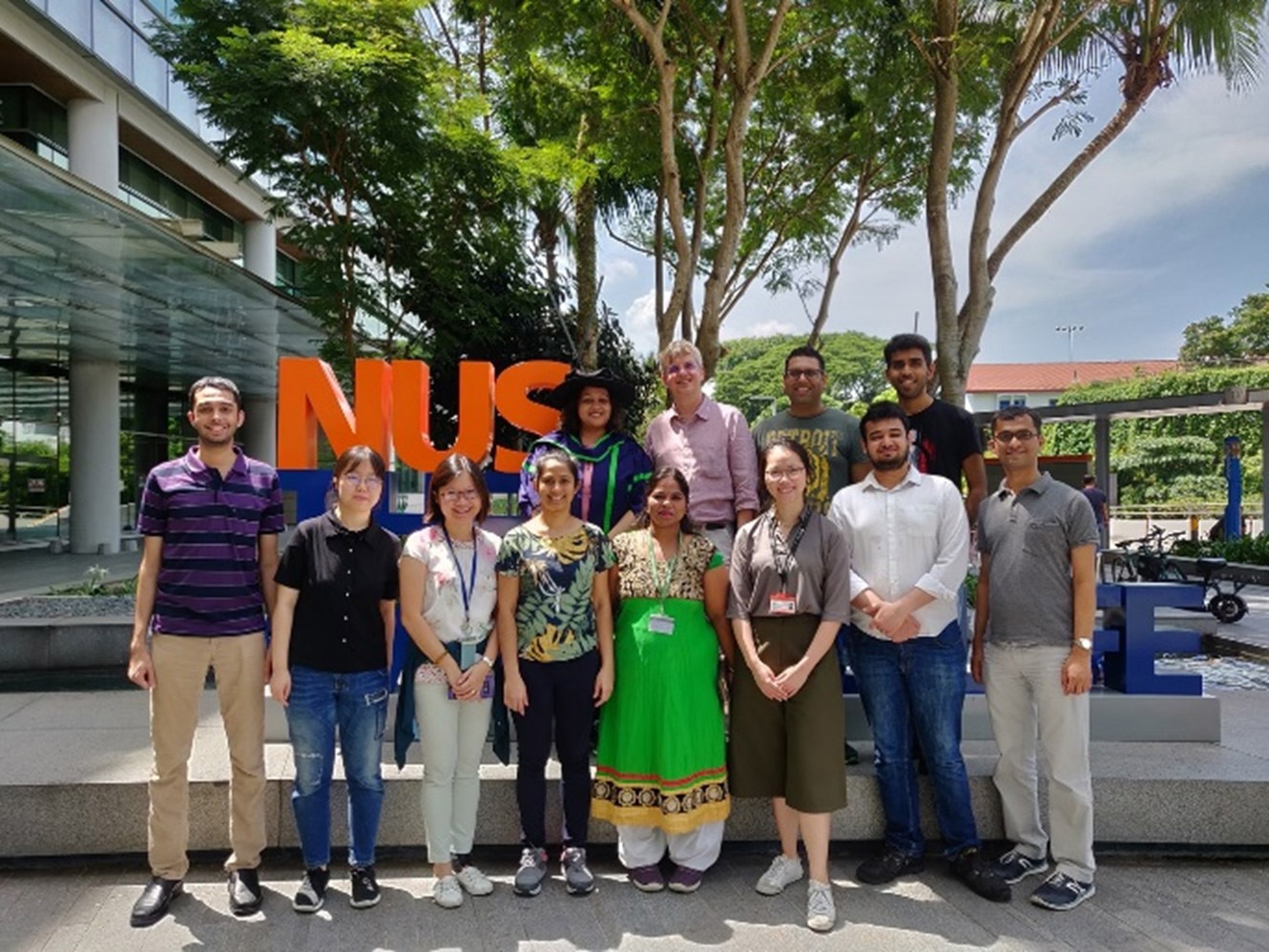
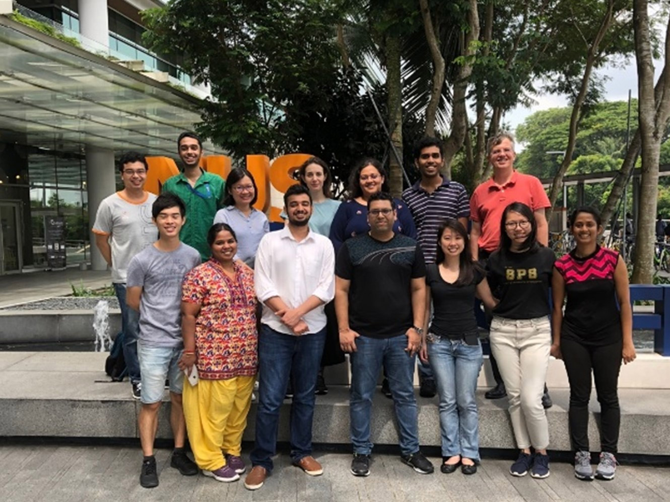
Above: Members of the Wohland Lab
Date: August 2021
Author: Daniel Aik, Jagadish Sankaran, Harikrushnan Balasubramanian, Centre for BioImaging Sciences, National University of Singapore
Category: Case Study
