Resources
 Part of the Oxford Instruments Group
Expand
Collapse
Part of the Oxford Instruments Group
Application Notes
Author: Dr Claudia Florindo
Published: 01 Mar 2022 · Last updated: 21 Mar 2022
This article presents an overview of scientific and technical challenges in cell cycle and cancer biology. We introduce the Andor Benchtop Confocal – BC43, the new instrument from Andor Technology. We show how BC43 overcomes the challenges in cell cycle & cancer biology research fields
Movie 1 – Live imaging of Cell division (Mitosis) observed with Andor Benchtop Confocal - BC43. Mammalian cells were imaged with BC43 using confocal imaging mode for over four hours. At each time point, four independent positions were imaged, and for each position, we acquired 3 channels and 15 Z stacks. The movie shows one of the 4 positions. Looking at the microtubules (Yellow) to the DNA (Cyan), the cell going in and out of mitosis can be observed through the duration of the experiment. (Cyan – DNA, Yellow - Microtubules, Red - Sir Actin). Image credits: Ines Baião-Santos, Álvaro Tavares – Universidade do Algarve, Claudia Florindo – Andor Technology.
BC43 is Andor’s new plug and play high-speed confocal system. BC43 is a benchtop confocal imager that delivers remarkable imaging excellence at a unique price. With the BC43, the user can choose different imaging modalities according to their experimental goals. Three imaging modalities are available and can be used simultaneously:
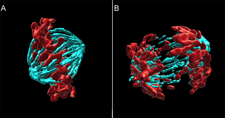
Figure 1 – Mammalian cell division observed with Andor benchtop confocal - BC43. Widefield Imaging modality was used to image fixed mammalian cells. A) metaphase cell, B) Anaphase cell. The image shows 2 of the 20 independent positions acquired in this experiment. We have acquired two independent channels for each position and covered a range of 15 mm. The deconvolution option was activated in the protocol. Images were further processed in Imaris, and we present the MIP of a surface rendered image. (Cyan - Microtubules, Red-DNA). Image credits Ines Baião-Santos, Álvaro Tavares – Universidade do Algarve, Claudia Florindo – Andor Technology.
BC43 is an ideal system for multiple life sciences applications including cell and cancer biology. The fast acquisition speeds cut down the imaging and processing time, boosting productivity without compromising image quality.
Andor Benchtop confocal delivers access to the 3D cell and tissue environment hundreds of micrometres deep into tissues. Furthermore, its gentle imaging of live cells and tissues for hours or days enables the visualisation of cell division (and cellular over-proliferation) within the tissues.
Importantly, all these imaging capabilities are packed into simple to use hardware and software integrated with easy acquisition workflows.
“It’s an outstanding microscope; it delivers remarkable image quality with a surprisingly fast learning curve. I found that young students doing short projects in the lab can get results almost immediately, which is something that I have never seen before in any confocal system.”
Professor Álvaro Tavares, group leader of the Cell Cycle and Cancer Biology Lab, Universidade do Algarve.
The main imaging challenges the cell cycle & cancer biology fields are summarised in the following table, along with the solutions offered by BC43 to overcome those challenges.
| Challenge | Solution |
| Live Imaging: Execute high-quality live imaging without phototoxicity or photobleaching. | Dual micro-lens multi-pinhole system with highly efficient excitation filter and detectors allow sensitive imaging, resulting in minimal phototoxicity and photobleaching. Live imaging of cells and tissues can be accomplished for periods over 24h. Widefield imaging option for thin, highly sensitive samples. |
| Fast Imaging: Accomplish live imaging of fast dynamic events. | Fast imaging speeds up to 44 frames per second (fps) either in confocal or widefield imaging modality. Fast imaging speeds are achieved with 10 ms exposure times (ensuring signal detection). |
| Thick Tissue Preparations: Image deep inside thick tissues. | Optimised pinhole spacing and size allow high background rejection, delivering a high signal to noise for images over the hundreds of micrometres range. |
| Large Tissues Imaging: Image through large samples, Image multiple live samples | Large Field of view (18.4 mm) allows increased productivity (fewer images needed to acquire the whole sample (1.84 mm Filed of view in the 10X objective). Integrated motorised stage and stitching. The full sample area can be captured and automatically stitched by selecting the stitching option on the acquisition protocol. |
| Analyse Multiple Samples (Increase productivity): Combine Multiposition with montage. -Fast confocal and widefield imaging. - Inline stitching and deconvolution. | Multiposition-montage option allows the acquisition of multiple sample areas in a single experiment – increase productivity. Multipoint confocal delivers confocal images as fast as 44 fps and 10X faster than regular point scanners. Increase productivity for both fixed and live imaging experiments. Protocol activated Inline deconvolution allows increasing images signal to noise ratio and resolution of the acquired images Protocol activated stitching delivers the full image result soon after acquisition. Both Stitching and deconvolution can be activated in the protocol. The full processed image, as well as the raw data, are delivered soon after acquisition. |
For more information on BC43, please read the article Introducing the new Andor Benchtop Confocal Microscope - BC43
Bibliography

© Oxford Instruments 2026
