Resources
 Part of the Oxford Instruments Group
Expand
Collapse
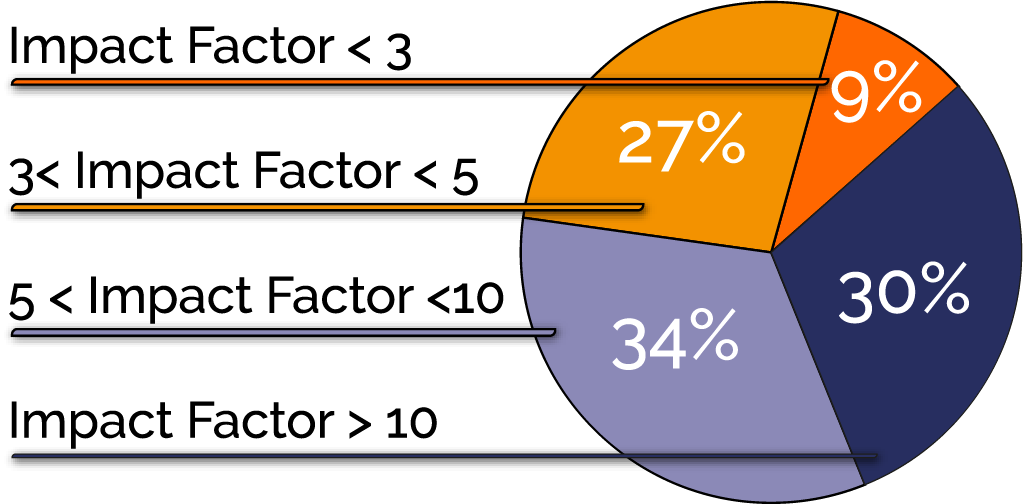
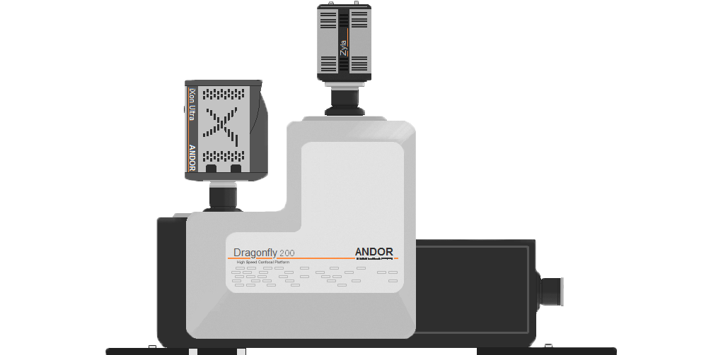
Since its launch, more than 200 papers have been published in high profile journals using Dragonfly. Dragonfly is a unique spinning disk confocal system that revolutionised the way imaging systems are perceived.
Before Dragonfly, there was no instrument compatible with live-cell imaging that could deliver deep-tissue imaging, as well as single-molecule localisation microscopy.


At Andor it matters to us that the products we develop make a real difference to our customers and contribute significantly to their research. We were born out of Queen’s University and have never forgotten our roots. It excites us that the Dragonfly platform is being widely used in papers published in high-impact journals like Nature, Neuron, Journal of Cell Biology and more.


64% of Dragonfly papers are published in journals with an impact factor of > 5
Dragonfly is much more than a high-speed confocal. TIRF, SRRF, and powerful laser-based widefield illumination can deliver super-resolved understanding of processes and place them in the context of whole tissues and organisms. Combined with Imaris, market leading 3D analysis software, our workflows address a broad range of disciplines with a single solution.
Disease Research

Clinical & Drug Studies

Technique Development

Membrane Dynamcs

Neuroscience

Mitochondria

Cell Division

Stem Cells

Actin

Centrosomes & Cilia

DNA Damage

We presented actual membrane trafficking events captured using Andor’s Super-Resolution Radial Fluctuation (SRRF-Stream) implemented via the highly sensitive iXon camera. Andor´s imaging technology offered new insights on the role of PI3-kinase in clathrin-mediated pinocytosis. Understanding this fundamental mechanism of vesicle trafficking could have implications for many diseases.
Cytokinesis is the physical separation of two cells that occurs after the completion of mitosis. In this webinar, we will present how we used a combination of optical techniques such as subcellular optogenetics, FRAP, TIRF, confocal imaging (Dragonfly) and SRRF-stream imaging to uncover the membrane dynamics during the final steps of cytokinesis.
At the recent 3rd birthday celebration of the Andor Dragonfly confocal system, Dr. Peter March (Bioimaging Facility at The University of Manchester), Biology's answer to Professor Brian Cox, took us on a humorous journey through the life of a Senior Experimental Officer at a growing Bioimaging Facility, including a brief history of Microscopes from the 17th century!
The Andor Learning Center hosts a wide range of tutorial videos, technical articles and webinars to guide you through the range of products for all your imaging needs. We have provided some links below which will get you started on some of our most recent uploads.

Dragonfly is an invaluable system for us and the busiest in our core facility. It’s been very helpful for our model organisms and organoid work, as well as being the first system in ESRIC which can do live super-resolution imaging.
Dr Ann Wheeler, Head of Advanced Imaging Resource, IGMM, Edinburgh

Dragonfly integrates speed of acquisition coupled with little or no photobleaching which has helped me immensely during my mitochondrial imaging. It is a wonderful system to perform both live and fixed cell imaging.
Rajarshi Chakrabarti, PhD, Research Associate at Geisel School of Medicine at Dartmouth