Resources
 Part of the Oxford Instruments Group
Expand
Collapse
Part of the Oxford Instruments Group
Challenge Background
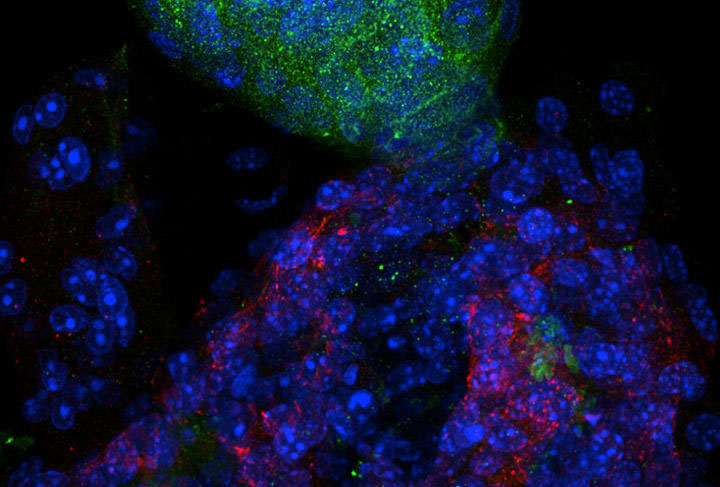
Organoids may be thought of as simplified versions of organs that have been produced in vitro usually from stem cells. They have a self-organised three dimensional structure and share many attributes with the corresponding organ. Organoid studies are proving ever more valuable since they are an effective way to closely recreate the physiology of various in vivo tissues and whole organs like liver or kidneys. Amongst the many areas that organoids have been used for are: preclinical models for cancer studies, screening of potential regeneration medicines, in the development of novel drugs and for drug efficacy testing. An example of how organoids can be used is given in the article Organoids in the study of liver development and cancer.
Imaging organoids effectively can be difficult as the large areas of cells that constitute the organoid under study need to be imaged in high resolution. Often this must be done in vivo and/or in three-dimensions to facilitate a more accurate understanding of the fundamental cellular processes we are interested in. The camera needs to have excellent sensitivity to be able to detect the low light levels that are inherent to the imaging techniques used such as spinning disk confocal laser microscopy (SDCLM) or light sheet microscopy (LSFM). In addition, the camera ideally should have a wide field of view so we can see as much of the subject under study in one view as possible.
Do you need the ultimate sensitivity? Check out the new iXon Life 888 EMCCD camera with single photon sensitivity – ideal for the most demanding low-light experiments.
Technology Solution
sCMOS cameras are known for their large sensor sizes and ability to run at high speeds which means that large areas of samples like organoids can be imaged in high resolution. The latest sCMOS cameras with back-illuminated technology are even more sensitive making them more effective at imaging under the low light regimes that are typical of experiments in this area.
Andor Camera Solutions for Study of Organoids
Andor highly recommend the Sona back-illuminated sCMOS camera for organoid studies in which sensitivity and field of view are of the upmost importance. Sona is perfectly suited to these studies due to the combination of sensitivity, field of view and speed. A near perfect 95% QE and low noise makes Sona highly sensitive. Sona 4.2B-11 provides a massive 32 mm Field of View from its 4.2 Megapixel sensor with 11 m pixels so that more of the organoid can be observed in a single image without stitching. Sona 4.2B-11 is also fast – 24 fps at full resolution (2048 x 2048) and 48 fps when high speed mode is used.
| Key Requirement | Organoids Studies Solution: Sona 4.2B-11 |
| High quality imaging even at low light levels | With 95% QE and low noise the signal to noise ratio can be optimized even at low excitation powers and low light levels. This means that you can keep fluorophore concentrations low and exposures as short as possible for more accurate cell physiology and reduced photo-toxicity. Result – Use low fluorophore concentrations and short exposures for more accurate cell physiology. |
| View the largest possible sample area | Sona 4.2B-11 features a massive 32 mm FOV from its 4.2 Megapixel (2048x2048) sensor with 11µm pixels. This is 62% larger than competing cameras using the same sensor. This allows larger areas of cells or whole organoids to be seen in one image, with less need for stitching. Result – View the largest sample area possible and accelerate experimental throughput. |
| View images with a wide dynamic range | Sona features Extended Dynamic Range technology (EDR) for an impressive 53000: 1 dynamic range. Demanding images with both weak and bright signals can be fully resolved in just one snap. Result – observe highly dynamic images in full detail in one snap. |
| See dynamic processes with excellent temporal resolution | Sona offers speeds of up to 48 fps at full frame (2048x2048), while ROIs boost speeds even further (e.g. 378 fps at 256x256). Sona lets you view the subtle yet significant changes in cell physiology. Result – follow dynamic processes and accelerate experimental throughput. |
| Quality and Longevity | Sona comes with Andor’s exclusive UltraVac™ vacuum sensor enclosure. The well proven permanent vacuum process is critical not only for cooling, but for protection of the back-illuminated sensor against moisture and condensates. Result – Sustained high performance, year after year. |

© Oxford Instruments 2026