Optically Detected Magnetic Resonance in Nitrogen Vacancy Ensemble
Author: Gerhard Wolff
Published: 01 Jan 2019 · Last updated: 24 May 2023
Quantum sensors offer a pathway to get better insight into processes at a small scale. Yet many quantum sensors can only be used under very specific conditions. The nitrogen vacancy centre (NV), due to its diamond host material and special electronic structure, offers a fully controllable single qubit system at ambient conditions, and is therefore an interesting candidate for investigating biological systems.
The incentive for a CCD setup compared to the more common tool of a confocal setup in the context of nitrogen vacancies is twofold. First the signal scales with the square root of the number of NV’s, which makes it interesting in the field of NMR [1]. Secondly by using multiple NV orientations, the strength and direction of external magnetic fields can be reconstructed [2].
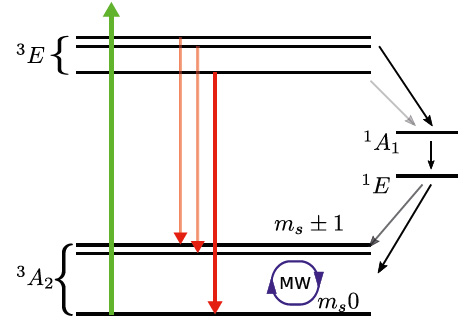
Fig. 1: Mechanism of the NV polarization under off resonant green laser light.
Experiment
In our experiments we typically use off resonant green laser light with a wavelength of 532 nm. The laser light is focused onto the back focal plane of an 80x Olympus immersion oil objective. This allows wide field illumination over a field of 30 μm and with that initialisation of the NV ensemble. At the heart of the initialisation and readout is the division in spin conserving and non-spin conserving decay processes. The spin conserving process occurs mostly through the transition from the |ms=0> excited to the ground state, whereas the |ms=±1, 0> excited states have a higher probability to decay over a metastable singlet state to the |ms=0> ground state. The nitrogen vacancy center can be manipulated by means of an external microwave field, which in our case is exerted by a 25 μm thick wire placed on top of the diamond sample.
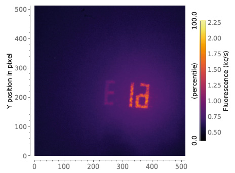
Fig. 2: Ion implanted marker consisting of NVs imaged using Andor’s EMCCD camera iXon ultra DU897-UCS-BV.
The marker in Fig. 2 depicts an ion implanted NV ensemble recorded with the Andor EMCCD camera iXon Ultra DU897-UCS-BV. To find the transition frequencies shifted due to the Zeeman splitting caused by an external magnetic field we continuously illuminate the NV ensemble for polarization and exert microwave fields with varying frequencies by means of an external signal generator.
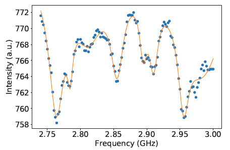
On resonance this reduces fluorescence as population is transferred from the |ms=0> to the |ms=±1> state as can be seen in Fig. 3. There we integrated the signal coming from the pixels in the marker and plotted them over the applied frequencies.
Fig. 3: ODMR peak structure originating from different NV orientations in diamond under an external magnetic field.
References
[1] Dominik B Bucher, David R Glenn, Junghyun Lee, Mikhail D Lukin, Hongkun Park, and Ronald L Walsworth. High resolution magnetic resonance spectroscopy using solid-state spins. arXiv preprint arXiv:1705.08887, 2017.
[2] D Le Sage, K Arai, DR Glenn, SJ DeVience, LM Pham, L Rahn-Lee, MD Lukin, A Yacoby, A Komeili, and RL Walsworth. Optical magnetic imaging of living cells. Nature, 496(7446):486, 2013.