Resources
 Part of the Oxford Instruments Group
Expand
Collapse
Part of the Oxford Instruments Group
Case Studies
Author: Andor
Published: 10 Feb 2021 · Last updated: 10 Feb 2021
The Endoplasmic Reticulum (ER), Golgi, endosomes, lysosomes, mitochondria and plasma membrane, all of which are membrane structures perform a variety of functions in eukaryotic cells. In order to accommodate these functions cellular membranes fold into various shapes, e.g. the filopodia protruding from the plasma membrane, the cristae of mitochondria, the cisternae of the Golgi, and the meshwork of the ER. These membranes undergo remodelling regularly and such dynamic processes play essential roles for maintaining the morphology and functions of these subcellular structures and organelles. Electron microscopy (EM) has been the technique of choice in the past for the study of these membranes at the nanometer-scale resolution. However, EM cannot be used to image live specimens and therefore dynamic information is lost. Super-resolution techniques such as STORM can overcome this difficulty by taking advantage of the use of photoswitchable probes and high precision localization of single molecules to surpass the diffraction limit.
Xiaowei Zhuang and her colleagues at Harvard University wanted to identify if commonly used fluorescent probes could also be used as photoswitchable probes for STORM imaging of living cells. They firstly investigated the use of carbocyanine dyes with long alkyl chains such as DiI, DiD, and DiR which have been widely used as labels for the plasma membrane. These dyes were found to exhibit photoswitching behaviour in the absence of an exogenous switching agent. Under 561-nm (for DiI), 657-nm (for DiD), or 752-nm (for DiR) illumination, these probes fluoresced and rapidly switched off to a dark state; the dark-state molecules could then be reactivated to the fluorescent state by 405-nm illumination. Per imaging frame each dye molecule emitted on average 790 photons. This reversible photoswitching behaviour allowed for the capture of STORM images of the plasma membrane on live hippocampal neurons. The fast switching rates of DiI meant a super-resolution image could be captured in 15 seconds or less. Images with a resolution of 40 nm were captured using Dil as the plasma membrane probe.
Three photoswitchable probes; Mito Tracker Orange/Red which are cationic rosamine dyes and Mito Tracker Deep Red which is a cationic carbocyanine were identified to capture STORM images of the fast dynamics of mitochondria. Similar to Dil, these dyes can be excited to fluoresce and turned off by 561 nm (Mito Tracker Orange/Red) or 657 nm (Mito Tracker Deep Red) illumination and reactivated by 405 nm illumination. All three dyes were used to capture live STORM images of the mitochondria in BS-C-1 cells. Because mitochondria tend to move fast in cells, the camera frame rate was increased to 900 Hz. Using these mitochondrial probes and this fast capture speed, mitochondrial fission and fusion intermediates were captured with excellent temporal resolution of about 1 sec.
Two BODIPY conjugated probes were identified to study the ER and lysosomes; ER-Tracker Red, a BODIPY TR conjugate which binds to potassium channels enriched in the ER and LysoTracker Red, a BODIPY 564/570 linked to a weak base that is highly selective for the acidic membrane of lysosomes. Again, similar to that of DiI it was discovered that both BODIPY TR and BODIPY 564/570 were capable of photoswitching in live cells. They could be imaged and switched off by 561-nm illumination and reactivated by 405-nm light.
The study revealed eight commercially available membrane probes each specific to the plasma membrane, mitochondria, the ER or lysosomes that can be used as photoswitchable probes for super-resolution fluorescence imaging. These probes readily label live cells with high probe densities allowing for very accurate localisation precision. Dynamic imaging of specific membrane structures was achieved in living cells with 30–60 nm spatial resolution at temporal resolutions down to 1–2 s. Due to the spectral properties of these probes two-colour super-resolution imaging was performed and ultra-structural dynamics of the plasma membrane, ER and the mitochondria were captured. These discoveries promises to reveal ultrastructural dynamics of organelles that are essential for cellular function.
Andor Solutions for Live STORM
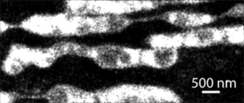
Live STORM image of Mitochondria
For this particular study iXon 860 was used for its extremely fast acquisition speed and sensitivity. The full range of iXon EMCCD cameras can be used for live STORM and in particular the iXon Ultra 897 with the “Optically Centred Crop Mode” which facilitates the capture of highly dynamic events such as mitochondrial fission and fusion. Facilitated by a fundamental redesign, the new iXon Ultra platform takes the popular back-illuminated 512 x 512 frame transfer sensor and overclocks readout to 17 MHz, pushing speed performance to an outstanding 56 fps (full frame), whilst maintaining quantitative stability throughout. The significant speed boost offered in the iXon Ultra 897 facilitates a new level of temporal resolution to be attained, ideal for speed challenged applications such as live super resolution techniques. Andor can also offer Zyla 4.2 for live STORM applications. Zyla 4.2 with the highest QE sCMOS sensor (> 72 %) can acquire extremely fast frame rates (100 fps at 4.2 MP, faster at smaller ROI’s) with very low noise (0.9 electrons) offering the sensitivity and speed required for super-resolution applications.

© Oxford Instruments 2026
