Resources
 Part of the Oxford Instruments Group
Expand
Collapse
Part of the Oxford Instruments Group
Application Notes
Author: Dr Alan Mullan and Dr Claudia Florindo
Published: 01 Feb 2020 · Last updated: 10 Feb 2021
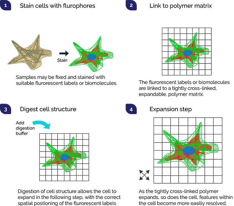
Like many of the developments in this list, Expansion Microscopy is another technique that enables improved resolution of cellular components that are central to cell function. Polymerization of a sample in a gel matrix expands the features, and now with more separation between them, features can be visualised using a conventional microscope. This novel approach is therefore broadly accessible to many labs for e.g. neuroscience, cancer and disease studies. It can readily be combined with other techniques like light sheet or super resolution methods, to further improve its scope. Key to this technique are the expansion steps. These steps are: addition of the fluorophore(s) of interest, linking the cell to the gel matrix, digestion of cell structure that would otherwise hold the cell together, and the proportional expansion of the sample from its original dimensions to the expanded size without distortion.
This technique is relatively recent, so its contribution to research is only just getting started. High sensitivity and large field of view sCMOS cameras such as the Zyla and Sona series are perfectly suited to studies using Expansion Microscopy.
References

© Oxford Instruments 2026
