Resources
 Part of the Oxford Instruments Group
Expand
Collapse
Part of the Oxford Instruments Group
Application Notes
Author: Gerald Cairns
Published: 01 May 2018 · Last updated: 19 Sep 2023
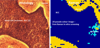
Raman techniques are being investigated for enabling fast diagnostic screening of various illnesses of the body to aid clinicians in providing more timely decisions in patient treatment plans. Cancer diagnosis is a prime example [1, 2, 3]. Currently the gold standard is biopsy followed by histology involving time consuming preparations and reviews of stained tissue samples. The trained pathologist assesses the samples for changes in cell structure, shape, etc to ascertain the state of health of the tissue sample. This in turn may infer changes in the molecular processes taking place within the cells.
Task at hand
Raman spectroscopy is a powerful technique for extracting information on the molecular content and processes within tissue cells and offers an extra dimension when determining the state of health of the cell. It involves the use of lasers to excite vibrational and rotational transitions within molecules. Inelastic scattered light is observed which reveals information on the energy levels associated with the particular molecular structure. Characteristic ‘fingerprint’ spectra are produced which enable the identification and concentration of the different molecules in the specimen. This is a form of molecular spectroscopy. Raman offers the advantage of not requiring sample preparation, such as sectioning and staining, and thus lending itself to quick in-vitro and in-vivo diagnosis.
Andor solutions
Andor provides technology for such Raman investigations in the form of high sensitivity, fast spectral rate, CCD and sCMOS detectors [7] along with high throughput dispersion spectrometers for the acquisition and analysis of Raman spectra. Some system combinations are summarised below.
MOHS surgery is an example where speed of diagnosing a tissue sample is of key benefit [4]. Samples are continuously assessed as part of the procedure to minimise the amount of healthy tissue removed during surgery (see picture above). Raman is also being considered for cases of in-vivo surgery [5, 6].
Background fluorescence from the excited tissue can be challenging when trying to measure the weak Raman emission, a problem which many researchers circumvent by working in the NIR region, for example, by using a laser at 785 nm to excite the molecules and observing the Stokes emission at longer wavelengths. Sensors optimised for the NIR are often chosen in these cases. Further information is found in the links to technical notes at the bottom.
Some very good combinations of instruments worth considering for fast screening:
| Camera | Spectrograph | Situation scenario | |
| 1 | iDus DU490A-BRDD | Kymera 328i | Good combination for wide range of application in VIS-NIR |
| 2 | Newton DU920P-BEX2-DD | Kymera 328i | High sensitivity performance with fast spectral rates in VIS-NIR |
| 3 | Newton EMCCD | Kymera 193i | High sensitivity, good throughput for low level signals and ultra-fast scanning |
| 4 | iVac | Kymera/Shamrock | For a good cost-effective ‘work-horse’ performance. Suited to OEM solutions |
| 5 | iDus DU491-LDC-DD | Kymera/Shamrock | Utilises LDC technology for sensitive measurements in the NIR region |
Links to useful articles for further more detailed information:

© Oxford Instruments 2026
