Resources
 Part of the Oxford Instruments Group
Expand
Collapse
Part of the Oxford Instruments Group
Application Notes
Author: D. Wu, A. F. Kukk
Published: 01 Sep 2022 · Last updated: 08 May 2024
Tags: modular-raman-spectroscopy-bio-matrials-fr
Raman spectroscopy is widely used in medical and life science research. As an important tool for learning about biological macromolecules, molecular-level information can be obtained by analyzing changes of the Raman spectral bands. β-carotene was dissolved in alcohol and isopropanol, injected into chicken skins for Raman measurements. Compared to fresh skin results, it was found that the increase in the intensity of the three carotenoid peaks is nonlinear, the decrease in the peak intensities of lipids and proteins is non-linear as well.
Raman spectroscopy is a spectroscopic technique based on the principle of Raman scattering, which is widely used in medical and life science research. Raman spectroscopy can detect the structure and composition of proteins, lipids, DNA, and other biological macromolecules in biological samples. When changes in the Raman spectral bands are analyzed, rich molecular-level information can be obtained, and changes in the structure or quantity of these biological macromolecules can also be obtained. Because of that, Raman spectroscopy is known as a “molecular fingerprint” [1].
Raman spectroscopy has very important applications in the field of biomedicine. For example, for the pigmented site on the skin, by identifying the changes of the peak intensities of proteins, lipids, and carotenoids on the Raman spectrum, physicians can judge whether it is a malignant tumor or not and monitor whether it is in an early or advanced stage, which provides a reference for the need for surgical treatment [2].
In this application note, we investigate the effect of carotene content on skin Raman spectra by injecting β-carotene into fresh chicken skins. β-carotene was dissolved in alcohol and isopropanol and injected into the skins for Raman measurements, the result is compared with that of fresh skin. In addition to the significant increase in peak intensities of carotenoid, we also found a non-linear decrease in peak intensities of proteins and lipids, suggesting that increasing levels of a certain chemical component in the skin can also affect other chemical components in different degrees. This factor cannot be ignored in future skin cancer Raman studies.
The Andor Kymera 193i-A-SIL spectrometer provided by Quantum Design is ideal for studying Raman spectroscopy of biological samples. It provides a wealth of custom functions and can be freely matched with different gratings to meet the needs of specific situations. With the Andor iDus DU416A-LDC-DD camera and a grating of 1200l/mm-600nm, the spectral resolution of 0.22 nm can be achieved under the excitation light wavelength of 532 nm, which is very suitable for Raman measurement of the animal epidermis. Three groups of chicken skins were removed from fresh chickens purchased on the market, one group was not treated as a control, and the other two groups were injected with 1 ml saturated β-carotene ethanol solution and isopropanol solution, respectively. Each sample was irradiated with a Q-switch 532 nm laser (20 Hz, 7 ns pulse duration) for 10 s at an energy of 1mJ and the Raman scattering signal was collected by the spectrometer.
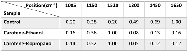
Table 1 intensities of typical β-carotene, lipid and protein peaks in different treated skin spectra
As can be seen in the table, with the absence of carotene injection, the amide-I band at 1650 cm-1, is about 4-5 times stronger than all carotenoids peaks (1005 cm-1, 1150 cm-1, 1520 cm-1), while the intensity of the amide-I band at 1650 cm-1 after injection is similar to that of 1005 cm-1, and is much lower than that of the other two carotenoid peaks. That is to say, the intensity of the three carotenoid peaks in the spectrum after β-carotene injection does not increase linearly, but the peak intensity at 1520 cm-1, increases the most, that at 1150 cm-1, increases less, while the intensity at 1005 cm-1, has the least increase. In addition, the reduction of peak intensities in the protein and lipid bands is also nonlinear. For example, the intensity at 1650 cm-1, is reduced by a factor of 6-8, that at 1450 cm-1, is reduced by a factor of around 5, and the intensity at 1300 cm-1, has the most drop, reaching 6-10 times.
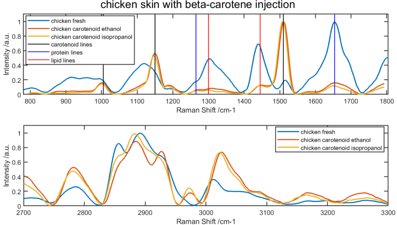
Figure 2: Chicken skin with and without beta-carotene injection, typical skin Raman bands have been marked in color lines
Raman spectroscopy has a very high potential in the biomedical field and is often regarded as a powerful tool for analyzing the molecular composition and structural changes in biological samples. In this application note, we measured and analyzed the Raman spectra of fresh chicken skins and chicken skins injected with β-carotene, and found that the increase of the intensity of the three carotenoid peaks was not linear, the intensities of other typical protein and lipid peaks decrease nonlinearly too. This indicates that, when the content of a certain chemical structure in an organism changes, it also has varying degrees of influence on other chemical components. In general, whether this effect holds for all organisms remains to be demonstrated, but this feature can be further used in the diagnosis of cancer by analyzing the difference in protein and lipid peak intensities in Raman spectra of diseased and normal skin to distinguish benign and malignant tumors and different stages of cancer.
References
Lyon L A, Keating C D, Fox A P, et al. Raman spectroscopy[J]. Analytical Chemistry, 1998, 70(12): 341-362.
Auner G W, Koya S K, Huang C, et al. Applications of Raman spectroscopy in cancer diagnosis[J]. Cancer and Metastasis Reviews, 2018, 37(4): 691- 717.
Contact:
Anatoly F. Kukk
Hannover Center for Optical Technologies, Leibniz University Hannover Nienburger Straße 17 D-30167, Hanover Germany
Phone: +49-511 762 14686
E-mail: anatoly.kukk@hot.uni-hannover.de

© Oxford Instruments 2026
