Resources
 Part of the Oxford Instruments Group
Expand
Collapse
Part of the Oxford Instruments Group
Application Notes
Author: Dr Alan Mullan
Published: 01 Feb 2023 · Last updated: 23 Mar 2023
Optogenetics has emerged as a powerful tool to activate, control and study individual cells within a wider network of neurons. As more photoactive proteins and techniques continue to be developed, researchers in fields of research far from neuroscience are finding ever more ways to avail of the expanding optogenetics toolset. A fundamental requirement for conducting these experiments is the precise control of illumination parameters over the specimen. Therefore, a means to deliver illumination of certain wavelengths of a certain power level, to certain features of a cell, or tissues at precisely the right time.
An additional requirement for some experiments is that illumination may be required across complex regions, and/or multiple regions simultaneously with precise time synchronization:
A further benefit of multi-region illumination is simply being able to run more experiments in parallel. Get more data and run more treatments, or greater statistical confidence in datasets.
There are two approaches for illumination available for use in optogenetics and optophysiology studies. The first of these is using a beam steering approach whereby a galvanometer can be used to steer the laser across a sample. This lets you target regions of interest, as you select and direct illumination beam of a certain size to them. However, it does not allow precise time alignment for illumination over multiple regions, being limited by nature of steering a single laser beam. Nor does it allow use of more complex illumination patterns and shapes found in biological specimens.
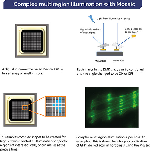
The other way to control the illumination of the sample is based on digital-mirror arrays. These devices are based around a DMD chip as would be found widely in modern video projectors. Each of the mirrors of these chips can be switched on to reflect light, or off (mirror angled to reflect light out of the image path). This technology makes it possible to define multiple regions of interest in whatever shape needed. Crucially illumination is delivered at each of these regions of interest at the exact same time. This digital-mirror array-based approach, if implemented in a suitable way coupled with a light source like LED or laser, is thus ideal for these applications that require illumination of multiple ROIs at the same time, as well as many others that need control over illumination region.
Mosaic is built around a DMD array specifically configured for optogenetics experiments. Multiple regions of interest are thus illuminated simultaneously and precisely as required. Complex user defined “masks” can easily be configured for user-created ROIs. This is simply not possible using a galvanometer-based laser steered approach.
Use Mosaic with your microscope and illumination source such as LED, laser, MicroPoint, or as part of a Dragonfly Confocal System and benefit from the suite of optogenetics techniques in your experiments.
| Feature | Benefit |
| DMD based illumination device | ✔ Allows simultaneous illumination across multiple ROIs with precise time synchronization ✔ Configure any shape of complexity of ROIs for illumination ✔ Protect sample outside of illuminated area from photodamage |
| Select from range of triggering and pattern sequence options | ✔ Range of triggering options to suit different experiments ✔ Cycle illumination patterns |
| Compatible with low power LED through to higher power laser sources such as MicroPoint | ✔ Perfect for bleaching or ablation at higher power densities ✔ Perform experiments that require lower illumination levels such as uncaging and photoactivation. |
| Match with upright or inverted Microscopes | ✔ Remarkable flexibility to configure for your specific experiments ✔ Have simultaneous imaging and photo-stimulation of the specimen |
| Range of UV-Vis imaging quality Epi illumination adapters available | ✔ Inserts between the epi-illuminator and trinocular head of upright microscopes ✔ Uses the epi-fluoresecent port of inverted microscopes ✔ Select excitation filters and/or beamsplitters to match your specific needs |

© Oxford Instruments 2026
