Resources
 Part of the Oxford Instruments Group
Expand
Collapse
Part of the Oxford Instruments Group
Application Notes
Author: Dr Alan Mullan and Dr Claudia Florindo
Published: 01 Feb 2020 · Last updated: 10 Feb 2021
Whether it’s with a Bessel beam, a Gaussian beam, an Airy beam or an Optical Lattice, Light Sheet Fluorescence Microscopy (LSFM) is now an established technique for imaging three dimensional biological samples e.g. tissues, organoids and model organisms like Zebrafish. LSFM provides an effective way to optically section the sample and minimise damage due to illumination, reject out of focus fluorescence and, image quickly and over extended periods of time. Although sample preparation takes extra consideration compared to other approaches, the technique is well suited to live cell imaging, proving very useful in developmental studies of drosophila, c.elegans or zebrafish. The different beam types provide different benefits making them suited to different sample types and there are many self-build and complete solutions available.
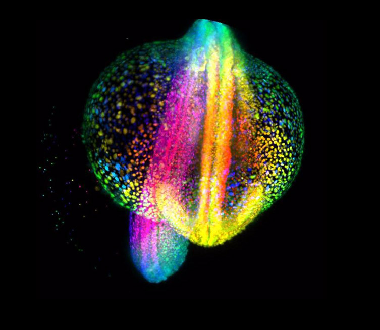
Light-sheet fluorescence microscopy imaging of Zebrafish embryo gastruliation from 4 to 18 hours post-fertilization, where each cell nucleus is labelled with GFP. Cells are color-coded for depth to visualisze how dynamic cell reorganization gives rise to the body axis of zebrafish. Image courtesy of Gopi Shah, Max Planck Institute of Molecular Cell Biology and Genetics, Dresden.
sCMOS cameras are a vital element of many LSFM set-ups as they have the required sensitivity, the imaging speed, and large sensor area that facilitates scanning large areas of the specimen at high speeds with minimal bleaching and high signal to noise.
Further Reading
References

© Oxford Instruments 2026
