Resources
 Part of the Oxford Instruments Group
Expand
Collapse
 Part of the Oxford Instruments Group
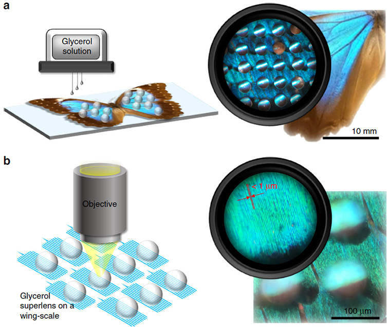
In recent work by Jia and co-workers at City University Hong Kong and the Shezen Academy of Robotics, glycerol superlenses were used to obtain super-resolution images of the intricate shingle structures on butterfly wings. Butterfly species studied included Morpho menelaus menelaus (M. m. menelaus) and Agrias beatifica beata (A. b. beata). A mixture of water and glycerol was prepared in a commercial liquid dispenser and deposited directly onto the substrate using a drop-on-demand preparation technique to fabricate the superlenses. The technique is outlined in Figure 1.

Caption Figure 1: Reproduced from Jai et al. Schematic depicting the printing of 3D superlenses directly onto the solid sample.
The glycerol superlenses in this work allowed features between approximately 100-200 nm to be resolved on the surface of the wings, resolving significantly more features than the microscope alone ~ 288 nm. The benefits of liquid superlenses over traditional techniques are that they are biocompatible, can be directly deposited onto the solid substrate and act as a relatively affordable route to super-resolution. Additionally, liquid lenses applied to a solid substrate in air can achieve superior resolution than solid microlens media. Liquid lenses offer increased resolution due to closer refractive index matching between the liquid lens (n = ~ 1.46) and the surrounding air, compared with solid microlenses such as barium titanate glass (n = ~1.9) and air. Moreover, liquid superlenses offer improved contact between the sample and microscope lens with the potential to develop other liquid solutions suitable for superlens preparation.
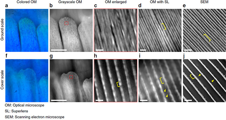
Jia et al. obtained super-resolution images using an optical microscope (OM) and a Zyla sCMOS camera with glycerol superlenses. Results were compared against images without superlenses taken using a Zyla sCMOS, an iPhone 7 Plus and a Scanning Electron Microscope. A selection of images obtained in the study are shown in Figure 2. Optical microscopy images taken with superlenses (SL) in d and i, demonstrate increased resolution and contrast when compared with images c and h where no superlens is used.

Caption Figure 2: Reproduced from Jia et al. Images of M. m. menelaus ventral wing scales. The Zyla 5.5 sCMOS camera from Andor was used to obtain images b-d and g-i using optical microscopy with a 100x (NA 0.9) objective. Whereas, images a and f were taken using an iPhone 7 Plus camera and e and j with an SEM. Scale bars: 50 μm for b and g; 2 μm for c–e and h–j.
Overall, Jia and co-worker's glycerol superlenses coupled with Andor’s Zyla sCMOS offer a route to cost effective super-resolution for solid substrates with rapid in-situ preparation of liquid microlenses directly onto the sample.
Alternative Routes to Super-Resolution
A family of super-resolution techniques are available to the life science researcher. Different techniques may be better suited to one application over another. Here are some links to a selection of resources on super-resolution available from our Andor Learning Centre, including articles on STORM, SIM and Andor’s SRRF-stream. Andor’s SRRF-Stream is an example of a camera-based means to achieve real-time live super resolution imaging.
References
