Plasmonic cavities are a powerful tool to enhance optical processes in nanoscale materials. The tightly confined optical fields at the junction between metallic nanoparticles can lead to an increase of the local light intensity by several orders of magnitude. This enables surface-enhanced Raman scattering, surface-enhanced infrared absorption, strong coupling, and the Purcell enhancement of light emission.
Here, we use plasmonic cavities to enhance photoluminescence (PL) upconversion. This is a process, in which light is emitted at a larger energy than the excitation laser. The PL upconversion can in general occur through two-photon excitation or through the extraction of thermal energy from the material. The latter process can even lead to a radiative cooling of the material, simply by shining light on it. PL upconversion occurs in many material platforms including rare-earth doped solids, semiconductor nanoparticles, and 2D materials. Especially in 2D materials the upconversion is usually weak because of a missing combination of energy levels for excitation and emission. In our recent work [1], we activated an excitation channel for PL upconversion in monolayer WSe2 by using the near-field coupling of plasmonic cavities to a spin-forbidden dark exciton.
Setup
We used a fully automated setup to characterize how the PL of monolayer WSe2 is enhanced by plasmonic nanocavities. The PL spectra were recorded with an Andor Newton 970 BVF EMCCD camera attached to an Andor Kymera 328i spectrometer with a 150 l/mm grating. The PL was excited and collected in a home-built dark-field optical microscope, with automated nano-positioning stages, an optical camera, and excitation with two cw lasers (633 nm and 785 nm) (Fig. 1). The nanocavities were identified by their dark-field scattering pattern using image recognition algorithms and centered to the measurement spot. Time traces of PL spectra were recorded on each nanocavity to monitor the stability of the PL and exclude damage. On each cavity, PL spectra were subsequently recorded with 633 nm and 785 nm laser excitation. The two lasers were combined with a dichroic beam splitter, toggled with shutters, and the respective notch filters in front of the spectrometer were switched with a motorized filter slider (Thorlabs). All instruments were interfaced with a home-written Python interface nplab (https://github.com/nanophotonics/nplab), making use of Andor’s software development kit. This enabled us to subsequently record the PL spectra of hundreds of individual plasmonic cavities.
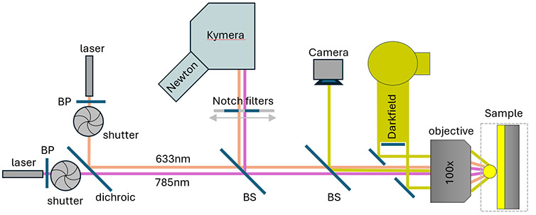
Figure 1 Schematic optical setup combining darkfield imaging with 633nm and 785nm laser excitation for single nanoparticle-on-mirror optical interrogation.
Results
We used plasmonic nanoparticle-on-mirror (NPoM) cavities to enhance the light emission of 2D materials (Fig. 2a). Au nanoparticles were drop-casted on an Au substrate that was covered with exfoliated flakes of monolayer WSe2. In this geometry, the 2D material defines a nanometre gap between the nanoparticles and the Au mirror. The plasmonic nanoparticles act as optical antennas confining light to this nanometre gap, which leads to a thousand-fold increase of the local light intensity.
The cavities were located through their dark field scattering patterns, which have the shape of horseshoes (Fig. 2b). When focusing the excitation laser beside a cavity, we observed weak light emission from WSe2 (Fig. 2b, left, dashed circle). This PL emission was strongly enhanced when centering the excitation laser on an NPoM cavity, which shows the Purcell enhancement by the plasmonic cavity (Fig. 2b, right). The enhancement was tightly located to the positions of the plasmonic nanocavities, with an up to 60-fold local increase of the PL intensity (Fig. 2c, d). PL spectra from a linescan across one of the cavities revealed that the light emission predominantly occured through upconverted PL that has a shorter wavelength than the 785 nm excitation laser (Fig. 2c).
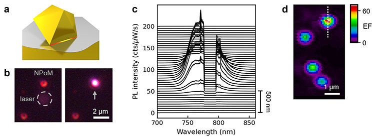
Figure 2 (a) Sketch of nanoparticle-on-mirror (NPoM) plasmonic cavity with decahedral-shaped nanoparticle. 2D material (grey) defines a nm gap between the nanoparticle and an Au substrate. (b) Optical dark-field + PL image of two NPoM cavities with excitation laser focused beside cavity (left) and on cavity (right). (c) PL spectra from linescan across NPoM cavity. Excitation laser lexc = 785 nm is blocked by notch filter. (d) Map of PL enhancement by four NPoM cavities. Line scan in (c) is indicated by dashed line. Panels (a), (c), and (d) are adapted from Ref. [1].
We explain this strong enhancement of PL upconversion through the interplay of dark and bright excitons in monolayer WSe2. The bright exciton (lbright = 750 nm) has an in-plane transition dipole, while the spin-forbidden dark exciton (ldark = 775 nm) has an out-of-plane transition dipole (Fig. 3a). The plasmonic near fields of NPoM cavities have, both, in- and out-of-plane electric-field components, which enables coupling to both exciton species. This is visible in a Stokes PL spectrum recorded with 633 nm laser excitation, i.e. excitation above the bandgap of WSe2, where we observed two emission peaks from the bright and dark excitons (Fig. 3b). A reference spectrum recorded beside the cavity showed weaker PL that is dominated by emission from the bright exciton (Fig. 3b, grey). This shows the Purcell enhancement of the PL emission by the plasmonic cavity and the activation of PL emission from the dark exciton, which was previously also observed by other groups with plasmonic cavities and tip-based approaches [2,3]. Using our fully-automated setup, we characterized the PL enhancement of 173 NPoM cavities. We found a systematically larger PL enhancement of the dark exciton than of the bright exciton (Fig. 3d, compare blue and red). This is because the plasmonic near fields in the nanogap of the NPoM cavity are mostly polarized out-of-plane, which leads to a preferential coupling to the dark exciton.
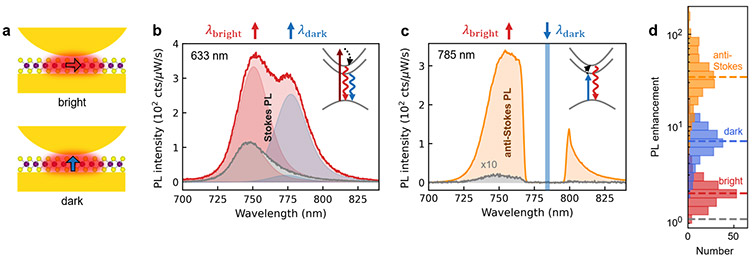
Figure 3 (a) Sketch of plasmonic coupling to in-plane transition dipole of bright exciton (top) and out-of-plane transition dipole of dark spin-forbidden exciton (bottom) in monolayer WSe2. (b) Stokes PL spectrum (lexc = 633 nm) of monolayer WSe2 enhanced by NPoM cavity (red) and recorded beside the NPoM cavity (grey). Two emission peaks occur from the bright (red) and dark (blue) excitons. (c) Anti-Stokes PL spectrum (lexc = 785 nm) enhanced by NPoM cavity (orange) and beside (grey). Laser excites dark exciton and emission occurs out of bright exciton at larger energy. See insets in (b) and (c) for energy diagrams. (d) Histogram of PL enhancement factors from measurements on 173 cavities, where enhancement of dark and bright exciton is obtained from Stokes emission spectra. Panels (b)-(d) are adapted from Ref. 1.
Interestingly, we observed a much larger enhancement of the PL emission when choosing 785 nm laser excitation (Fig. 3c,d, orange). In this configuration the excitation laser is near resonant with the dark exciton and light emission occurs predominantly out of the bright exciton at higher energy (Fig. 3c, anti-Stokes PL). This PL upconversion was almost absent beside the plasmonic cavities, which shows that the plasmonic cavities activate this process (Fig. 3c). Statistically, we observed a 10 to 100-fold enhancement of the PL upconversion, which is an order of magnitude larger than the enhancement of the Stokes PL (Fig. 3d, orange). When correlating the enhancement factors, we found that cavities which efficiently couple to the dark exciton also provided larger PL upconversion. The dark exciton thus serves as an excitation channel for PL upconversion, that is activated here by the plasmonic cavities.
In conclusion, we showed that plasmonic nanoparticle-on-mirror cavities activate PL upconversion in monolayer WSe2. This was accomplished through near-field coupling to a dark exciton, which served as an excitation channel, while emission occurred out of the bright exciton at higher energy. The simple fabrication protocol of the plasmonic cavities paves the way for large-area substrates, that may find application for nanoscale radiative cooling of 2D materials, anti-Stokes lasing, and radiative engineering of excitons. For these measurements the long wavelength studied makes it important that there is low dark counts, low read noise and a high quantum efficiency for the system. This makes the highly configurable Kymera spectrometer and the low noise Newton EMCCD suitable choices for these types of experiments.
References
[1] Mueller et al., Nat. Commun. 14, 5726 (2023), DOI: https://doi.org/10.1038/s41467-023-41401-8
[2] Lo et al. Nano Lett. 22, 1915–1921 (2022). DOI: https://doi.org/10.1021/acs.nanolett.1c04360
[3] Park et al. Nat. Nanotechnol. 13, 59-64 (2018). DOI: https://doi.org/10.1038/s41565-017-0003-0

