Stability - delivers reliable performance for long-term imaging experiments.
Resources
 Part of the Oxford Instruments Group

Fusion, developed by Oxford Instruments, introduces a new era in imaging software for confocal microscopy. Designed to perfectly complement our high-performance hardware, Fusion delivers a unified and highly adaptable platform for controlling confocal imaging systems: Benchtop-BC43 and Dragonfly, in addition to photo-stimulation devices: Mosaic.
Request Trial License Upgrade Fusion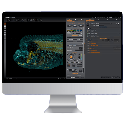
Fusion is designed to prioritise user needs, flexibility, and simplicity. It enables both benchtop microscopes and Dragonfly systems to be operated with minimal training, allowing efficient execution of complex, multidimensional experiments.
Fusion streamlines imaging by unifying acquisition, visualisation, and data handling in one intuitive platform. Its guided workflows and adaptive interface support users at any level, helping them move quickly from setup to high‑quality results. With smart automation and responsive performance, Fusion delivers consistent outputs while minimising setup time.

Stability - delivers reliable performance for long-term imaging experiments.

Ease of Use - features an accelerated learning curve for rapid user adoption.

Flexibility - supports a wide range of applications, e.g multidimensional experiments, poststimulation and others

Image Analysis – For advanced processing, analysis workflows, and data quantification, the Fusion software is seamlessly integrated with Imaris.

Modular - adapts the software to user requirements and upgrades as needs evolve.

Super-Resolution - compatible with super-resolution protocols, including SMLM and SRRF-Stream+.

Hardware Integration – BC43 benchtop confocal, Dragonfly, and Mosaic are all operated using Fusion software.

Image Processing - Integrates more than essential image processing tools required during acquisition, e.g., stitching and through-series analysis.
Fusion modules are designed for both simple and complex protocols, prioritizing productivity and ease of use. Please review each module to see how its features can enhance your image acquisition.
Click on the + to learn more about Fusion features.
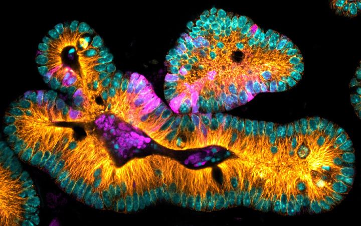
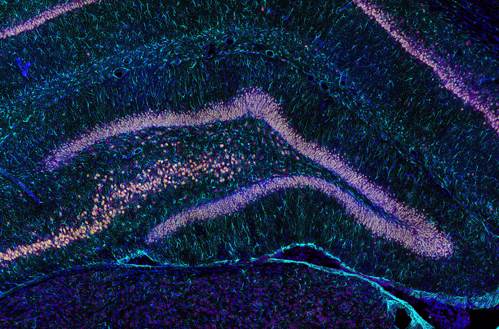
Image Legend: Mouse Intestinal Organoid expressing LGR5-GFP to visualize stem cells (magenta) microtubules (orange) and DNA (cyan). Image courtesy of Nicole Dawney from the Bergstralh Lab and High Content Imaging Core, University of Rochester.
Deeper Insight, Better Data
*1 Piezo available in Dragonfly Fusion only
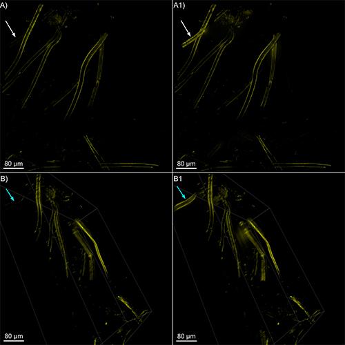
Image Legend – The Deep Z feature in Fusion reveals hidden structures at depth
A Z‑stack was acquired (A, B), and the Deep Z feature was applied (A1, B1). In panels A and B, the laser power was kept constant at 6%. In panels A1 and B1, the laser power started at 6%and increased exponentially up to 30%. Deep Z activation occurred at approximately 80 µm depth, after which the laser power ramped progressively to 30%.
A total of 231 stacks were acquired, corresponding to a 137‑µm imaging range. Structures that become visible due to the increasing laser power are indicated by the white arrows in A), A1)(MIP) and by the blue arrows B), B1) (orthogonal view).
Immediate experimental feedback
Grasp the dynamics of biology
Translate cell behaviours into biological insights by imaging dynamic processes in living cells.
*Available in Dragonfly only
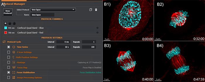
Figure Legend – Time-lapse setup and results
(A) Easy time‑lapse setup: select the time interval and number of repeats. Focus stabilization can be activated for higher positional accuracy. The quad‑band filter enable seven faster image acquisition. (B) Mammalian cell division sequence: (B1) Prophase, (B2)Metaphase, (B3) Anaphase, (B4) Telophase. Microtubules are shown in red, and DNA in cyan.
Stay sharp. Stay confident. Stay in focus.
Fusion control hardware-based autofocus maintains sample focus throughout the experiment. This feature is critical for live imaging assays, multi-well plate imaging, large fixed samples, multi-position imaging, and multi-tile imaging.
Reliable. Accurate. Always in focus
Multi-position Imaging of Mammalian Cells with BC43.
At each time point, four positions were captured in parallel, each with three channels and 15 Z‑stacks. Shown side‑by‑side, the video highlights how the Focus Seek & Lock technology keeps every position perfectly in focus—even during rapid multi‑site navigation. Image credits: Ines Baião‑Santos, Álvaro Tavares (Universidade do Algarve); Claudia Florindo (Oxford Instruments).
Expand your imaging capabilities
Configure multiple independent acquisition experiments and automate acquisition control for external devices.
Note – This is an advanced feature, and its use requires an understanding of Python coding
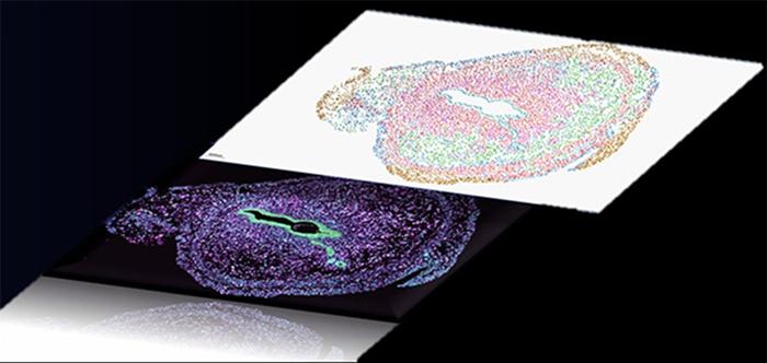
Spatial omics graphic representation
With the power of our REST API, users can orchestrate complex workflows by linking multiple protocols and external devices. Comprehensive gene-expression atlases can be generated by coordinating our spinning-disk confocal systems (Dragonfly or BC43) with automated microfluidic triggering.
Image Legend: Mouse Intestinal Organoid expressing LGR5-GFP to visualize stem cells (magenta) microtubules (orange) and DNA (cyan). Image courtesy of Nicole Dawney from the Bergstralh Lab and High Content Imaging Core, University of Rochester.
Effortless Sample Overview
See your entire sample instantly. With one click, our smart spiral montage delivers a complete, high-quality overview—no missed areas, no duplicate tiles.
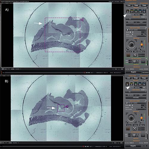
Image Legend – Overview of sample navigation & spiral montage
A) A 2×3 spiral montage was acquired using the 2× objective. The navigation icon (arrow), the 2× objective selection (arrowhead), and the montage definition (green rectangle) can be observed. B) When switching to the 10×objective (arrowhead), the field of view is automatically adjusted (arrow).
See Beyond the Field of View
Unlock the complete perspective of your sample with seamless, high‑coverage imaging.
Irregular montage new - Imaging That Adapts to Your Sample
NEW – Unlockhigher productivity by combining irregular montage with multiple positions
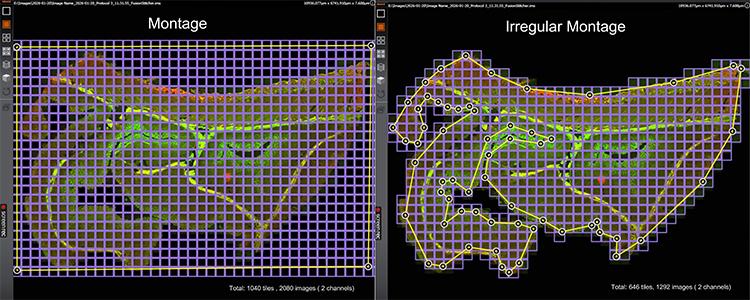
Figure Legend – Irregular montage increases productivity
This example compares a standard montage (left) with an irregular montage on the BC43 system (right). While the standard montage requires 1,040 tiles (2,080 images, 2 channels), the irregular montage reduces this to 646 tiles (1,292 images) by imaging only relevant sample areas.
The result is approximately 2× higher productivity for 2D imaging, with even greater gains for z-stacks and multi-channel acquisitions. For the same 3D sample, image count was reduced from 510,120 to 288,288, significantly decreasing acquisition time, data storage, and processing requirements.
In conclusion: Irregular montage acquisition captures only relevant sample regions, maximizing imaging efficiency while maintaining resolution and sample coverage.
Increase Productivity Through Higher Sample Throughput
Boost the efficiency of every experiment and extract more insight from each run.
1. More Output per Experiment - Deliver greater insight from every run without increasing workload.
2. Focus on What Matters - Acquire only the most relevant cells or sample regions to maximise efficiency.
3. Scale Further with Montage - Combine targeted acquisition with montage imaging for even higher coverage and context.
NEW – Unlock higher productivity by combining irregular montage with multiple positions.
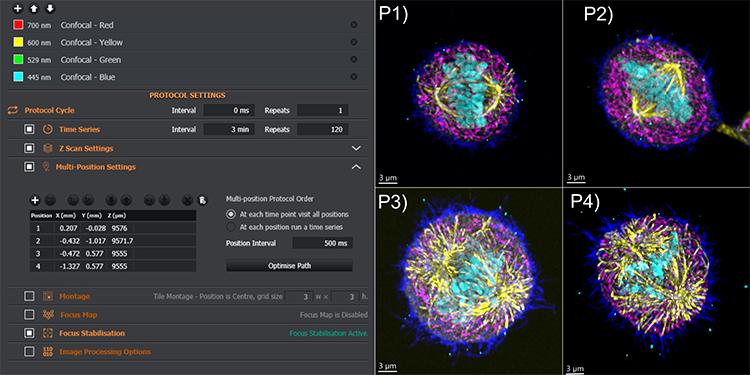
Multiple positions increase experimental throughput
In this time‑lapse, multichannel experiment, four positions were acquired to track mitotic cells, which can be rare depending on the tissue and cell line. The protocol included four confocal channels, time‑lapse, Z‑stack, and multi‑position acquisition with active focus stabilisation. Montage acquisition can also be added to further boost throughput.P1–P4 (right panels) show the four mitotic cells. Colour coding: dark blue – actin, pink – mitochondria, yellow – microtubules, cyan – DNA.
Redefine Throughput in Multi-Well Assays
Efficiently image and analyse multiple experimental conditions, reducing turnaround time and increasing throughput.
NEW – Unlockhigher productivity by combining irregular montage with multiple positions
One click well selection
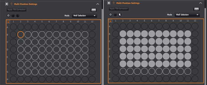
Navigate with well plate view or table view
All protocol combinations are possible.
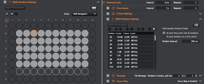
Keep every field in focus
Enables correction of tilt and sample unevenness. Can be used in multi-position protocols.
Supports robust acquisition of uneven and complex samples.
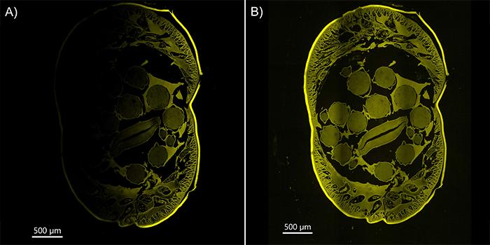
Figure Legend – Focus Map Correction of Slide Tilt and Sample Unevenness
Twelve tiles of the Ascaris sample were acquired. (A) Due to tilt in the slide, the left side of the image is not well focused and appears less visible. (B) After applying the Fusion Focus Map to the tilted Ascaris sample, both the left and right regions of the specimen appear sharply in focus.
Image Legend: Mouse Intestinal Organoid expressing LGR5-GFP to visualize stem cells (magenta) microtubules (orange) and DNA (cyan). Image courtesy of Nicole Dawney from the Bergstralh Lab and High Content Imaging Core, University of Rochester.
Illuminate Function, Frame by Frame
An integrated, precise, and flexible photostimulation workflow for advanced functional imaging.
Compatibility: Dragonfly + Mosaic systems.
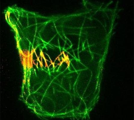
Perform Precise Photostimulation
Take precise control of photostimulation delivery. Target areas of tissues, individual cells or intracellular features. Gain insights into processes within living cells with powerful techniques such as photoactivation, FRAP, uncaging and optogenetics.
Simple, and accurate B-TIRF control.
B‑TIRF (Borealis TIRF) brings precision TIRF imaging to every user by simplifying a traditionally complex technique. With effortless setup and smart control, it delivers consistent, high-contrast and uniform illumination for demanding single-molecule and membrane-level studies.
SMLM or SRFF-Stream
Single‑molecule localization microscopy (SMLM) or push‑button super‑resolution—whichever fits your workflow.
SMLM – Single‑Molecule Localization Microscopy
(SMLM available for dragonfly only)
SRRF‑Stream – One‑Click Super‑Resolution
Achieves resolutions down to 140 nm
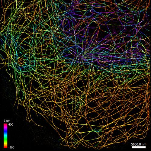
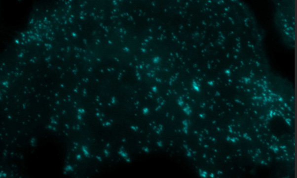
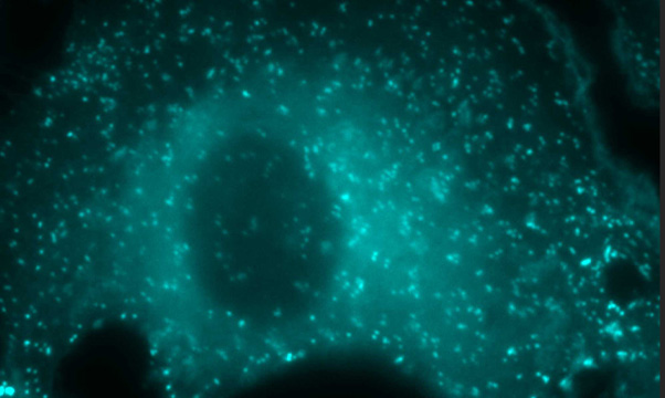
Microtubules in a cell imaged using Single Molecule Localization Microscopy
A 10,000 time‑point series was acquired to capture the stochastic emission of Cy3 fluorophores. The data were rendered, drift‑corrected, and subsequently localized, achieving a median localization precision of ~13 nm. Image acquired with the Andor Dragonfly spinning disk confocal. Data courtesy of Felix Rivera‑Molina, Yale University.
Image Legend: Mouse Intestinal Organoid expressing LGR5-GFP to visualize stem cells (magenta) microtubules (orange) and DNA (cyan). Image courtesy of Nicole Dawney from the Bergstralh Lab and High Content Imaging Core, University of Rochester.
See the big picture by visualising the cells within the context.
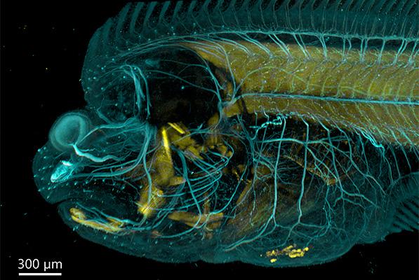
Figure – 3D stitching provides a full overview of the sample
Flat fish imaged on the BC43 using a montage. Six tiles were acquired to compose the image, covering a Z‑range of 554 µm. The 3D image was stitched using the Fusion option activated in the protocol.
(Image credits: Marco Campinho, Universidade do Algarve; Claudia Florindo, Andor Technology)
Sharper images, more detail, improved resolution.
ClearViewTM deconvolution enhances image clarity by removing haze and improving resolution.
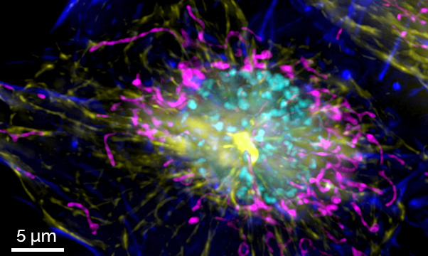
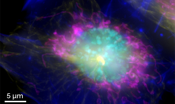
Enhanced resolution and contrast with ClearView™ deconvolution.
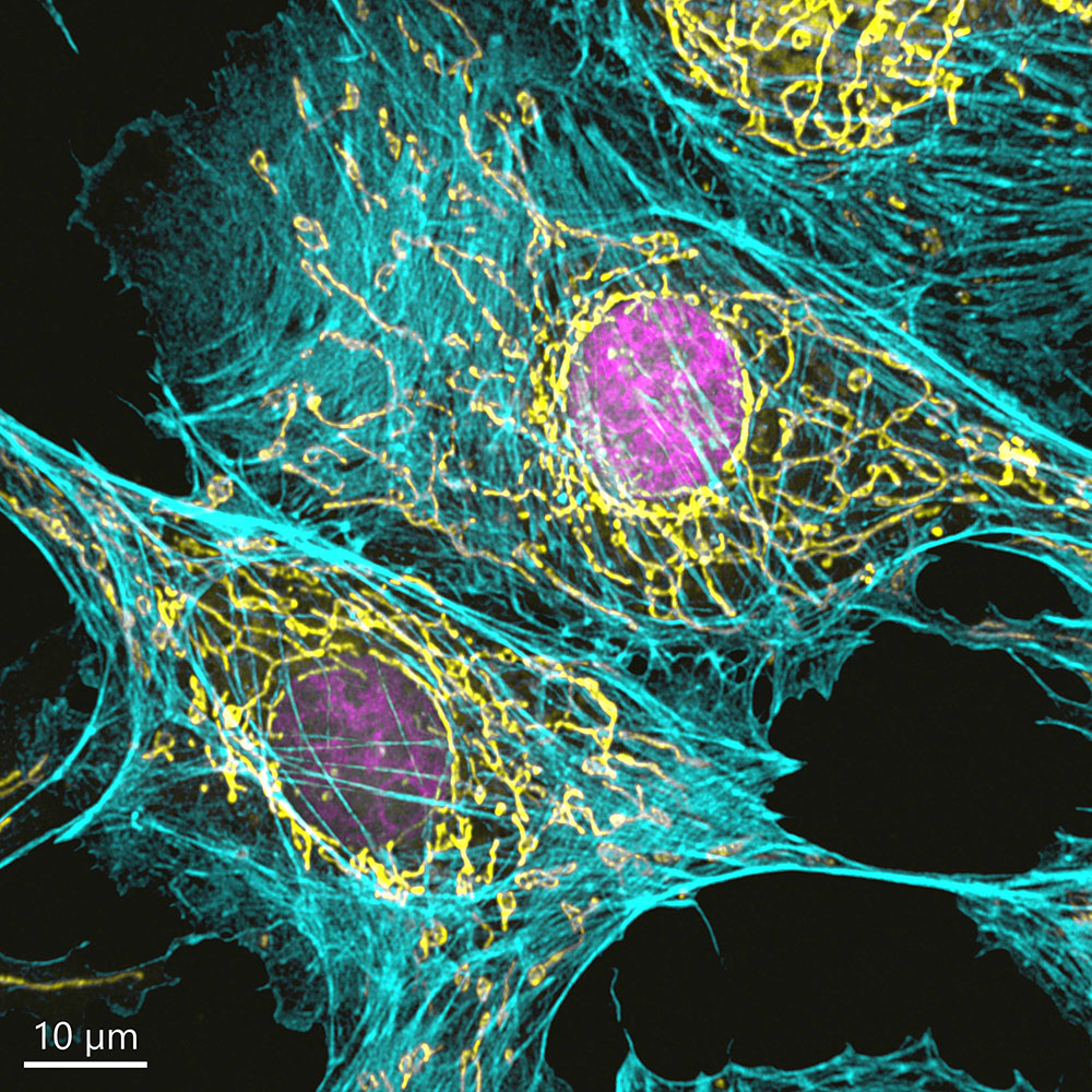
Widefield images of a mammalian cell acquired on the BC43 system demonstrate the improvement in contrast and spatial resolution achieved through ClearView™ deconvolution. Left: Widefield image , right: deconvolved image. Deconvolution was activated in the protocol. Fluorescent labels: actin (dark blue), microtubules (yellow), mitochondria (pink), and DNA (cyan). Image credit: Claudia Florindo, Oxford Instruments.
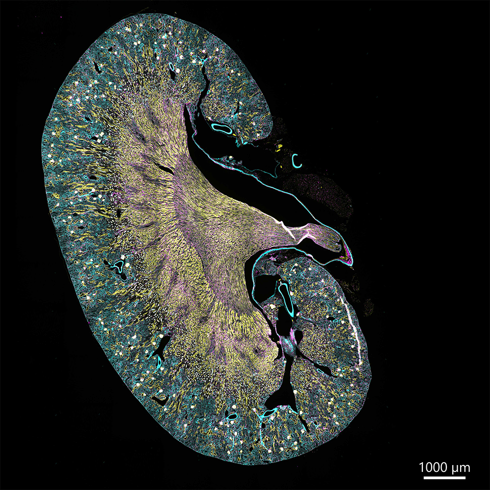
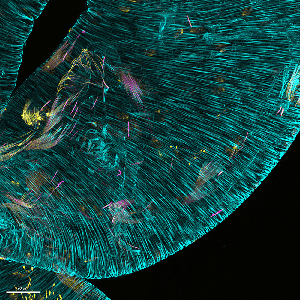
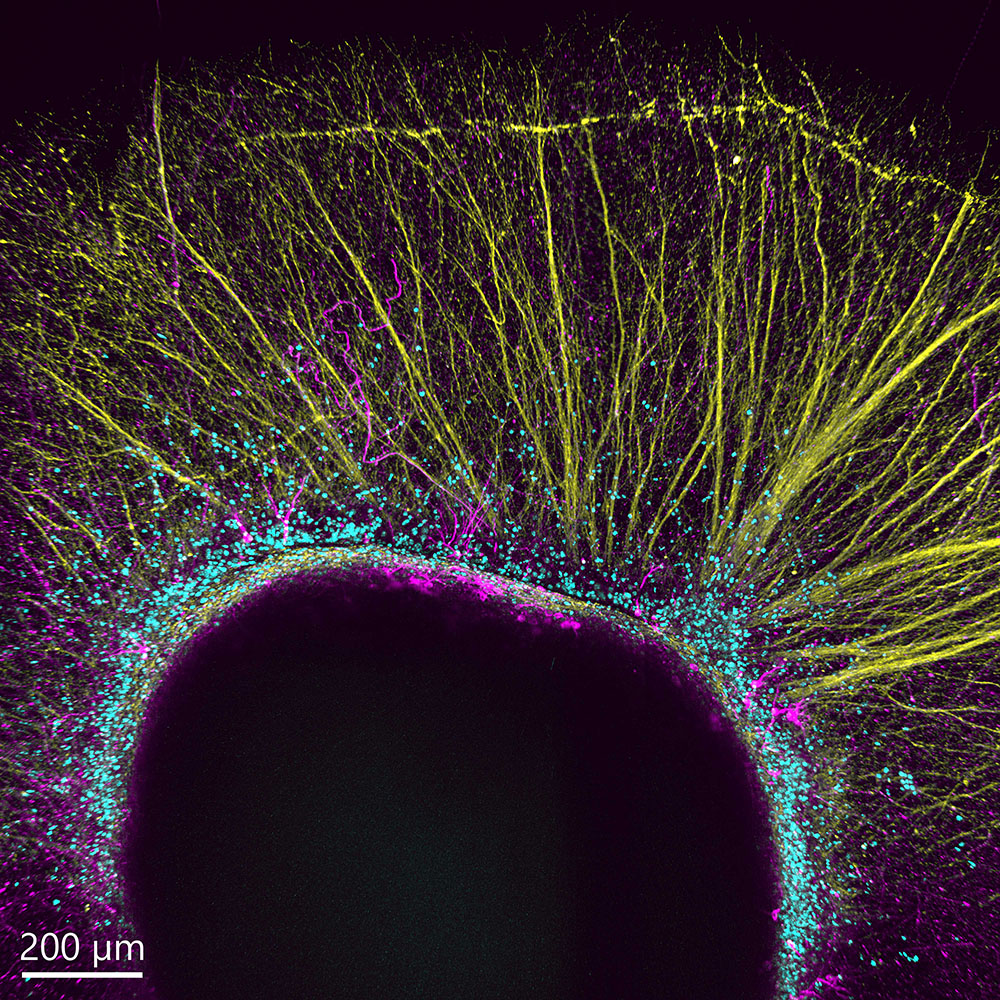
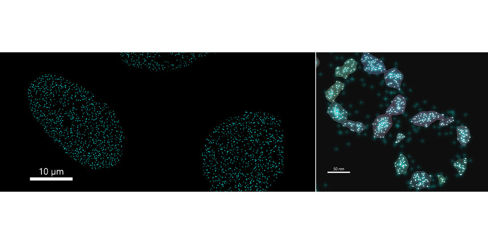
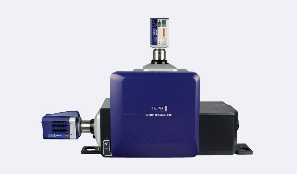
Dragonfly is a high-speed confocal platform that delivers exceptional images from subcellular structures to whole organisms with outstanding speed and sensitivity. It effortlessly supports live-cell, large model organism and SMLM imaging in a single, versatile system.
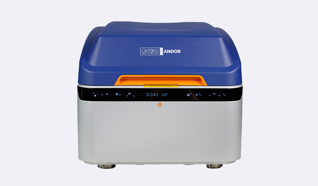
BC43 fuses entry-level simplicity with high-end performance in one compact benchtop system. It delivers 2D/3D widefield, confocal and push-button super-resolution. High-speed confocal boosts productivity, backed by an industry-leading certified quality program.
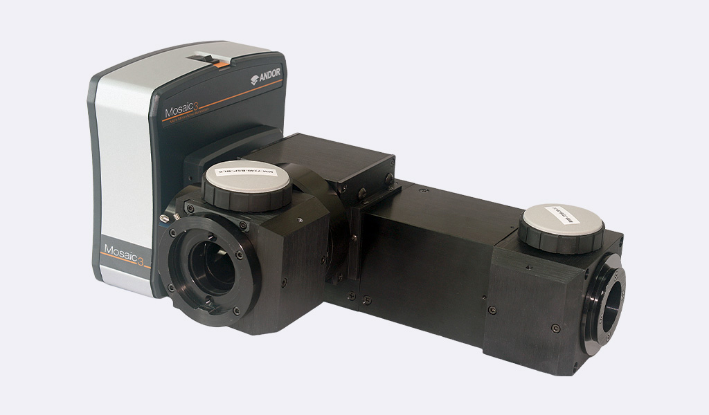
Mosaic lets you get more from your research with precise, targeted sample illumination using digital micromirror technology. It unlocks powerful photostimulation experiments, including optogenetics, photoconversion, uncaging and much more.

© Oxford Instruments 2026