Resources
 Part of the Oxford Instruments Group
Expand
Collapse
Part of the Oxford Instruments Group
Introduction
The correlation of functional response to structural changes remains a challenge for in situ and/or in-operando studies on highly localized anisotrophic phenomena. Therefore, a combined investigation of novel materials functional properties, such as photoluminescence, together with their structural properties, e.g. by X-ray diffraction (XRD) analysis is of high interest.
As an example for the performance of the system, we measured monolayers of transition-metal-dichalcogenides (TMDCs). The TMDCs family presents a native bandgap ranging from the ultraviolet to the infrared, a rather large in-plane electrical carrier mobility (up to 200 cm² V-1 s-1), strong light-matter interaction, and strong exciton-binding energies up to 1 eV at room temperature, which makes them ideal candidates for optoelectronic and photonic applications. [1]
In addition, the band structure in WSe2 shows a transition from an indirect band-gap in bulk to direct one for a single monolayer. As a consequence, WSe2 monolayer flakes exhibit bright luminescence properties. [2]
Experimental configuration
In the center of the system stands a six-circle diffractometer that allows a full strain characterization of crystalline specimen by recoding three dimensional reciprocal space maps for a set of Bragg reflections. Therefore, a collimated highly brilliant X-ray beam impinges on the sample. By rocking the sample around the Bragg condition, the strain state of the sample is investigated.
Simultaneously, the incident X-ray beam excites optical luminescence that is emitted by the sample, as depicted in Fig. 1. The optical collection system aims to increase the photon yield and minimize signal losses by making use of a collimating optics and a glass fiber to transport the light into the spectrometer.
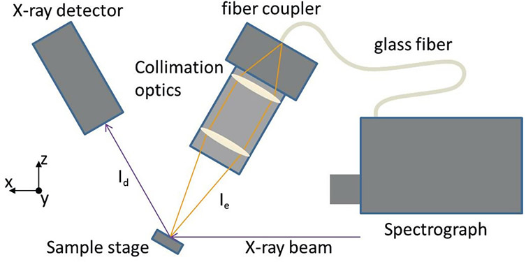
Figure 1: Schematic of the XEOL combined XRD configuration, indicating the emitted signal (Ie) and diffracted signal (Id) while the sample is mounted onto a six-circle diffractometer, and inclined by ~30 deg.
With a protective silver coating on all spectrograph mirrors, the highest reflectivity is achieved for a wavelength range from 400 nm to 1000 nm. A turret holding three different gratings, 300 l/mm, 600 l/mm and 1200 l/mm allows sufficient flexibility from wide range, overview spectra, to highly resolved spectra, respectively. The Andor multicore quartz glass fiber offers a core diameter of 200 μm (NA = 0.22) with an operation window from UV to VIS.
Sample preparation and first measurements
The measurements with the described collection system were first carried out at beamline P08 of the German Electron Synchrotron (DESY) in Hamburg. The portable set-up of the optical collection system allows for simple adaptation to different experimental end stations and a quick exchange. The 2D material system was excited with a monochromatic 15 keV X-ray beam.
The sample was produced by mechanical exfoliation to obtain flakes of WSe2, with sizes in the order of (5 x 10) μm². Such flakes were transferred to patterned Si (111) substrates with Au markers to identify individual flakes. An x-ray beam focused down to a size of (3 x 30) μm2 allowed to excite optical luminescence from a single WSe2 Monolayer, as seen in the inlay of Fig. 2. A full spectrum was recorded with an integration time of 100s in single photon counting mode and applying background correction (Fig. 2.)
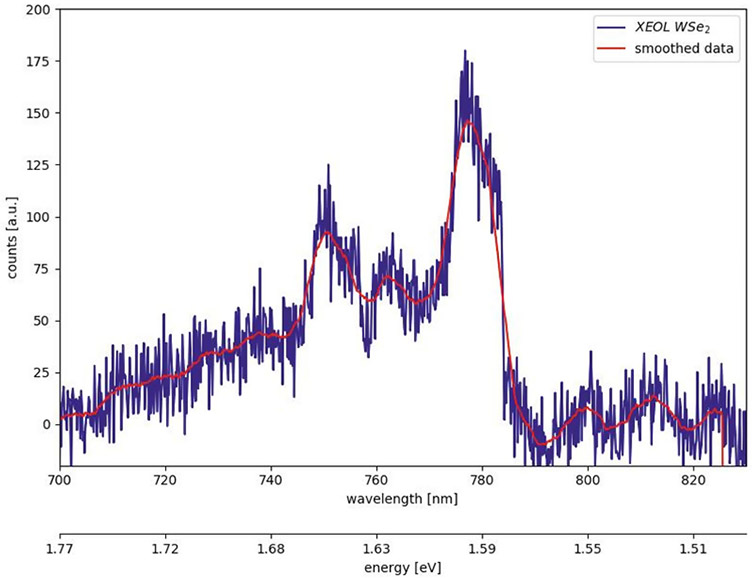
Figure 2: XEOL spectra from a single WSe2 Monolayer at RT. The inlay shows the specimen next to a neighboring Au marker, with a length of 20 μm, under an optical microscope.
Conclusion
XRD provides a model free insight to structural configurations with an exceptional flexibility for in-situ and in-operando measurements. At the same time the specimens’ functional response can be probed at the very same location and state.
The Andor iVac DR316B-LDC-DD CCD detector mounted on a Shamrock SR-303i-B-SIL spectrograph allows detecting full spectra from a single monolayer of WSe2, excited by highly brilliant X-rays. The combination of the individual components makes this system highly flexible for user-operation in a wavelength range from 400 nm to 1000 nm and can be used for the characterization of different material systems.
References

© Oxford Instruments 2026