Resources
 Part of the Oxford Instruments Group
Expand
Collapse
Part of the Oxford Instruments Group
Application Notes
Author: Andor
Evolution of atomic and molecular structure
In recent years a new method has been developed to study the time evolution of atomic and molecular structure on the time scale of 100 femtosecond (1fs=10-15s), which is the typical time scale for atomic vibration. This method will give new insights of the temporal evolution of physical, chemical and biological processes on the atomic scale. New developments, such as new X-ray sources, femtosecond lasers, and X-ray optics, were essential for this study. But without new detector development in the keV-photon range such experiments are not possible. A combination of a toroidally bent crystal optics with a CCD camera can provide the simultaneous measurement of transient crystal diffraction curves [1].
Time-Resolved X-ray diffraction using a pulsed femtosecond X-ray source at Institut für Optik und Quantenelektronik, in Jena
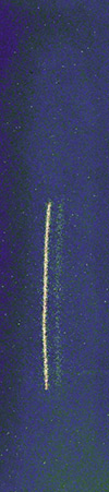
Fig. 2a.
A typical set up for Time-Resolved diffraction is shown in Fig. 1. The interaction of short intense laser pulses (t =100 fs, I >1015 W/ cm2) with solid matter creates a thin dense plasma layer where electrons can be efficient accelerated to keV or even MeV energies. Such electrons can produce short X-ray pulses when interacting with a solid. The X-ray source is slightly larger than the laser focus, typically produced some tens of micrometers. Intense line radiation from the laser based X-ray source, like Kα lines, are being focussed with toroidally bent crystals onto the samples which are investigated. Then a spherical monochromatic wave is falling on the sample. The diffracted X-ray signal from the sample (Fig. 2a) is recorded by an Andor back-illuminated, deep depletion X-ray CCD camera, a DX420-BR-DD with 1024 x 255 pixels, providing the rocking curve in the case of a single crystal sample (Fig. 2b). Excitation of the sample by a second laser pulse with a certain delay before the X-ray probe pulse, allows researchers to follow the temporal response of the diffraction signal by varying the delay of both pulses [2].
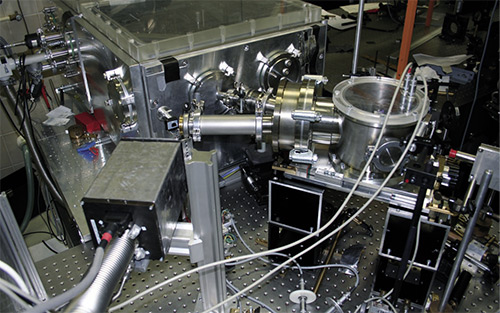
Fig. 1. Time-Resolved X-ray diffraction setup
Properties of X-ray CCD cameras
Important properties of back-illuminated deep depletion CCDs have the following advantages compared to other X-ray detectors [3]:
Application for Time-Resolved experiments
Time-Resolved experiments require significant properties of the detectors to record successfully reasonable data. One of the most important parameters for these experiments is the time average X-ray photon flux. A high detection efficiency reduces the extremely high cost for a high average power of the femtosecond laser driving the X-ray source.
X-ray CCDs are applied in various steps of these experiments:
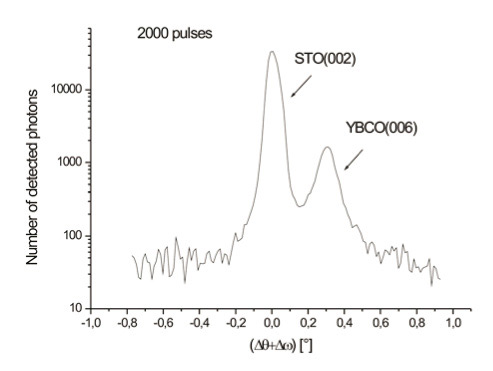
Fig. 2 (b). A single crystal rocking curve
Advantages for data analysis using back-illuminated deep depletion X-ray CCD cameras are:
With thanks to:
I. Uschmann, Institut für Optik und Quantenelektronik, Friedrich-Schiller-Universität Jena, Germany
1. Ch. Rischel, et al., Nature 390, 490 (1997).
2. A. Morak, et al. , Phys. Stat. Sol., No. pssb.200642387, (2006).
3. F. Zamponi, et al., Rev. Sci. Instrum., 76, 116101 (2005).

© Oxford Instruments 2026
