Resources
 Part of the Oxford Instruments Group
Expand
Collapse
Part of the Oxford Instruments Group
Case Studies
Author: Alan Mullan
Published: 01 Dec 2019 · Last updated: 10 Feb 2021
Challenge Background
The plasma membrane is a relatively thin yet important interface zone at which many processes that are critical to the cell occur. It is important not only for the internal processes and the actin cytoskeleton structure, but also for cell fate differentiation. The phospholipid bilayer acts as both a barrier and a point for interaction with the external environment. Proteins embedded within the plasma membrane such as transmembrane proteins are involved in cell adhesion, cell secretions and cell to cell communications. Study of these processes are providing insights into their roles within various neurodevelopmental disorders such as Attention Deficit/hyperactivity Disorder (ADHD) and Autism Spectrum as well as disorders of metabolism and aging.
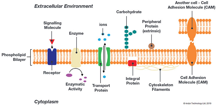
A simplified illustration of components of the plasma membrane. The plasma membrane is the site at which many critical cellular processes occur.
The study of the myriad of processes at the plasma membrane interface poses several challenges in terms of how to image processes and structures effectively. Techniques that are typically used to provide the necessary resolution and elimination of out of focus light such as TIRF (Total Internal Reflectance Fluorescence), light-sheet, SIM (structured illuminated Microscopy) or spinning disk confocal impose a low light regime making high sensitivity detectors essential. In addition, the imaging camera must be capable of a wide field of view so we can see as much of the subject under study in one view as possible. Some processes are dynamic in nature meaning high speed imaging is required to capture and resolve the temporal information.
Technology Solution
The latest back-illuminated sCMOS cameras provide exceptional sensitivity making them suitable for imaging samples that have low fluorophore signal levels, or when using imaging modalities that are inherently low light. As with previous generations of sCMOS cameras they feature low noise, large sensor sizes and an ability to run at high frame rates.
Andor Camera Solutions for Plasma Membrane Studies
Andor highly recommend the Sona back-illuminated sCMOS cameras for plasma membrane studies. Sona offers the perfect combination of high sensitivity, exceptional field of view and speed. A near perfect 95% QE and low noise means that the Sona is highly sensitive. Sona 4.2B-11 provides a massive 32 mm field of view so more can be observed in a single image. The 11 µm pixel is perfectly suited to resolving features in full clarify at high magnification. Sona 4.2B-6 is fast – up to 74 fps at full resolution (2048 x 2048) in full range (16-bit), and regions of interest can be used to boost speeds significantly higher e.g. 587 fps at 256 x 256.
| Key Requirement | Plasma Membrane Studies Solution: Sona 4.2B-11 and Sona 4.2B-6 |
| Detect small changes in fluorophore intensity | Sona is perfect for detecting small changes in signal intensity of fluorescent labels at the plasma membrane. With 95% QE and the deepest cooling, the lowest possible noise is achieved, making Sona the most sensitive sCMOS camera available. Fluorophore levels and exposures can therefore be kept as low as possible for more accurate physiology and reduced photo-toxicity effects. Result – Use low fluorophore and exposures for more accurate cell physiology. |
| View the largest possible sample area | Sona 4.2B-11 features a massive 32 mm FOV from its 4.2 Megapixel (2048x2048) sensor with 11µm pixels. This is 62% larger than competing cameras using the same sensor and over 114% larger than cameras restricted to a standard C-mount format. This means more image information in each snap. Result – View the largest possible sample area and accelerate experimental throughput. |
| Resolve the fine detail at the plasma membrane | Sona 4.2B-11 has a 11 µm pixel, which is perfectly matched for resolving the full detail of intricate structures and processes under high magnifications such as x100. Result – Resolve the full detail at high magnification. Sona 4.2B-6 has a 6.5 µm pixel, that allows superb spatial resolution at the widely used 40x and 60x magnification. |
| View dynamic events with excellent temporal resolution | Sona 4.2B-6 allows you to view processes such as endo and exocytosis with excellent temporal resolution. With speeds of up to 74 fps at full frame (2048x2048) , ROIs boost speeds even further (e.g. 587 fps at 256x256). Sona lets you view the subtle yet significant changes occurring at the plasma membrane. Result – observe changes in dynamic aspects of the plasma membrane. |
| Quality and Longevity | Sona comes with Andor’s exclusive UltraVacTM vacuum sensor enclosure. The well proven permanent vacuum process is critical not only for cooling, but for protection of the back-illuminated sensor against moisture and condensates. Result – Sustained high performance, year after year. |

© Oxford Instruments 2026
