Resources
 Part of the Oxford Instruments Group
Expand
Collapse
 Part of the Oxford Instruments Group
X-ray sources have improved significantly in recent years in terms of photon beam quality, energy resolution and number of photons. A high photon consumption method is called Resonant Inelastic X-ray Scattering (RIXS).
This technique is element, site and orbital specific as well as sensitive to the chemical environment and has become a valuable tool to investigate soft matter among others. An energy dispersive spectrometer is required to perform this experiment. The spectrometer consists of an optical element, which focuses the incoming fluorescence on a 2D detector.

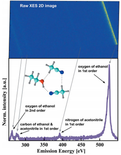
Fig. 1: The emission signal from three different elements (carbon, nitrogen and oxygen) are recorded at one detector position.
In our case, we used a varied line spacing (VLS) grating as the X-ray optic and a back illuminated Andor iKon-L CCD camera for direct X-ray detection. It offers a 2048 x 2048 pixel sensor with a pixel size of 13.5 μm. Compared to other available 2D X-ray detectors, the CCD provides high spatial resolution, which is an important part for the overall energy resolution of the spectrometer, and thus resolves spectral signatures.
Additionally, the CCD camera is user friendly and the cooling system can go down to -70 °C with air cooling, which is favorable for the signal to noise ratio. The large size of the CCD can fully exploit the VLS grating characteristics, which leads to the detection of a large energy range. The big chip can also cover a broad energy range.
As shown in Fig. 1, the CCD detector can cover up an energy range of ~300 eV, which includes the emission lines of three elements, in this case carbon, nitrogen and oxygen, while maintaining a sufficient energy resolution. This is an advantage because the experiment can be performed efficiently and contains more information about the electronic structure.
References
Author: Z. Yin, S. Techert, FS-SCS, Deutsches Elektronen-Synchrotron (DESY), Hamburg
Category: Application Note
