Resources
 Part of the Oxford Instruments Group
Expand
Collapse
Part of the Oxford Instruments Group
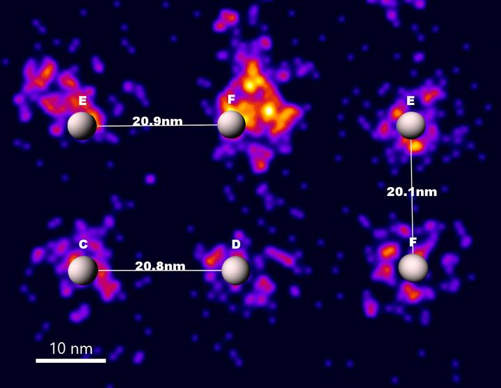
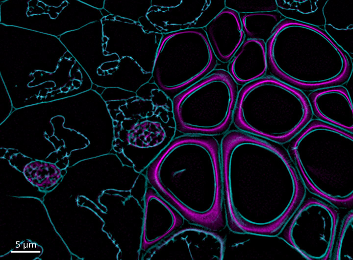
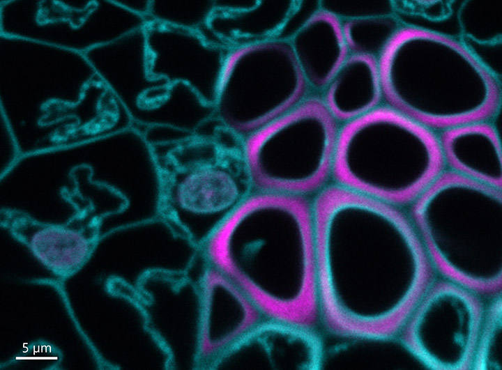
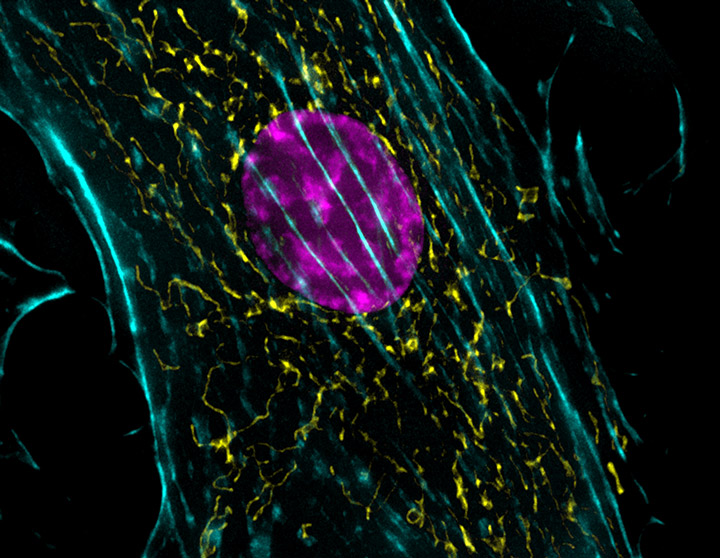
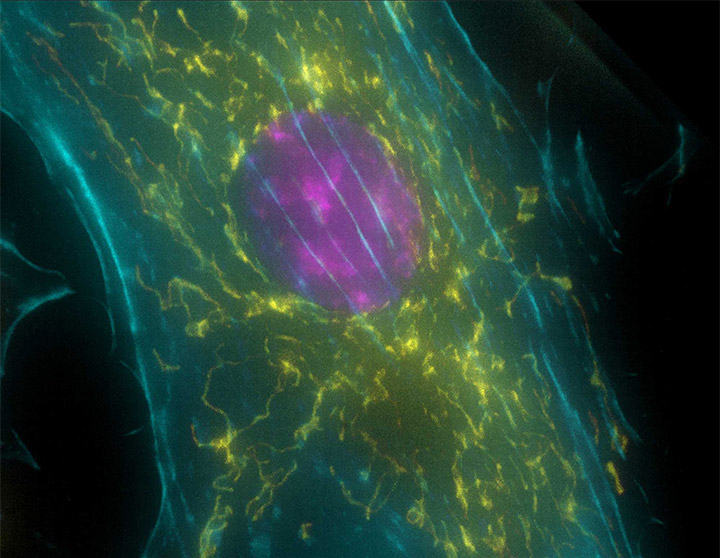
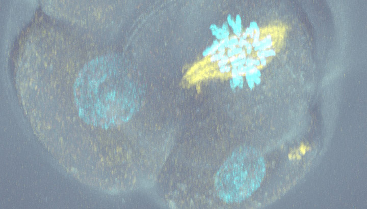
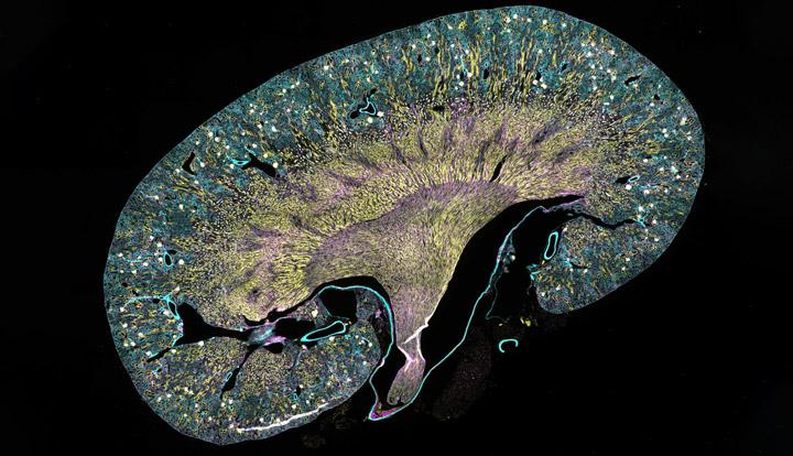
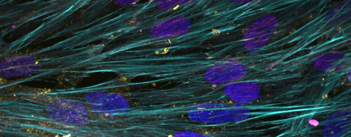
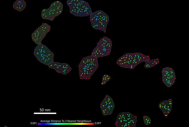
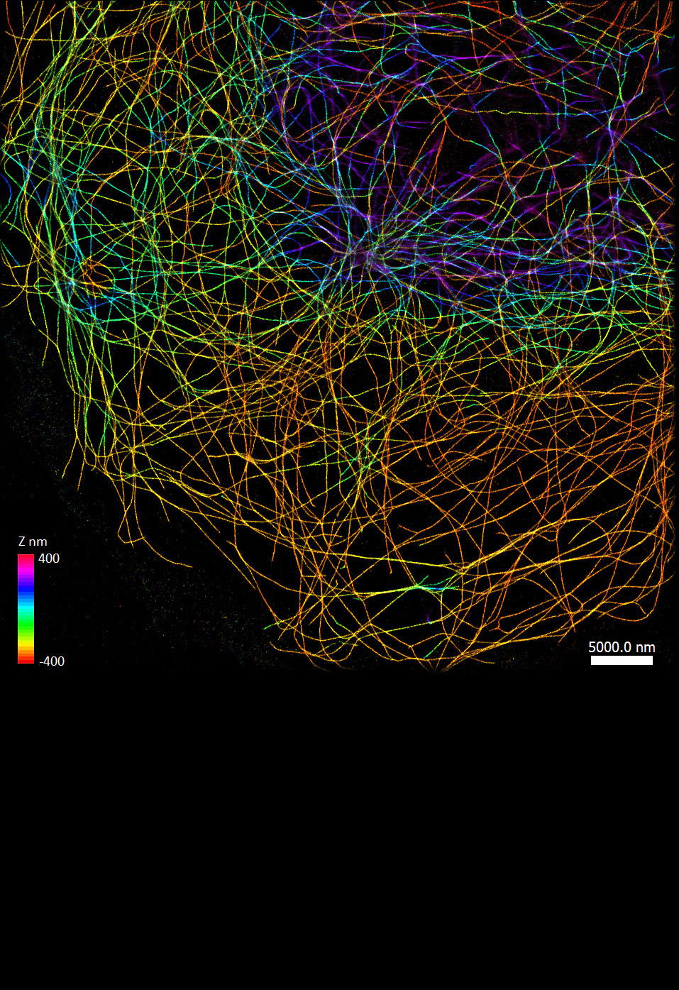
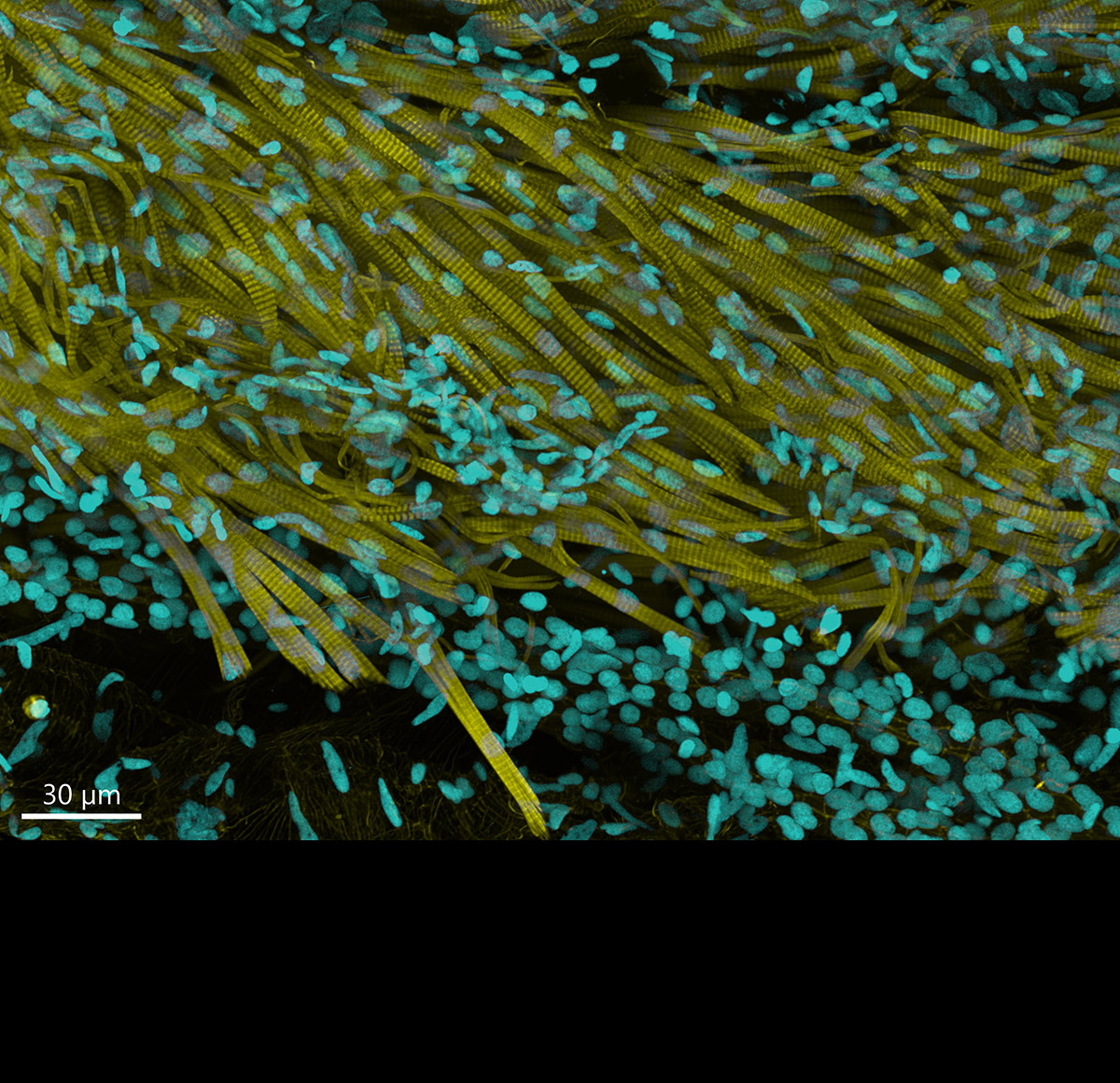
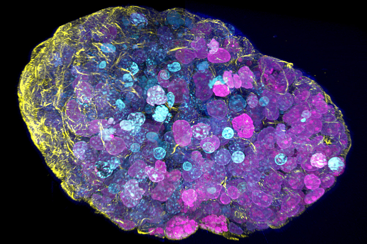
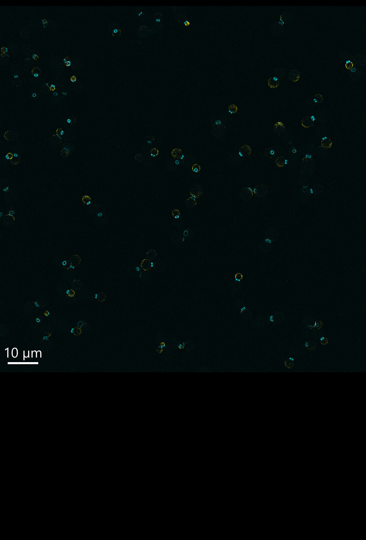
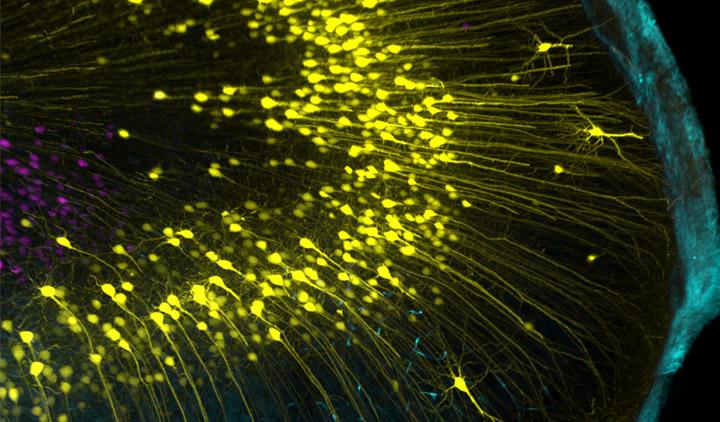
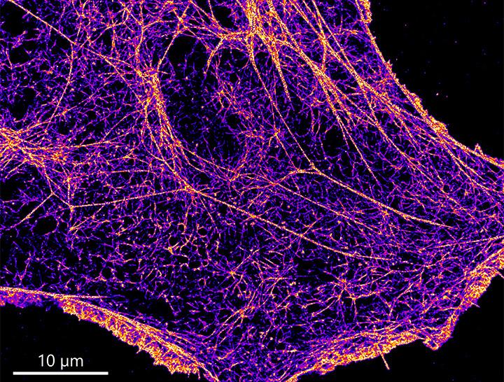
The Dragonfly provides high-quality confocal images across scales, from the subcellular level to entire organisms, and significantly increases productivity. It combines speed and sensitivity, enabling researchers to observe dynamic events and live organisms for extended periods. The B-TIRF and super-resolution modules reveal finer details, such as viral infection dynamics and chromatin or organelle ultrastructure.
Each Dragonfly high-speed confocal includes Fusion software for effortless image acquisition and Imaris, the market leader in image analysis software.The Dragonfly 600 is the flagship model. It delivers a full-featured system and allows for effortless cross-scale imaging.
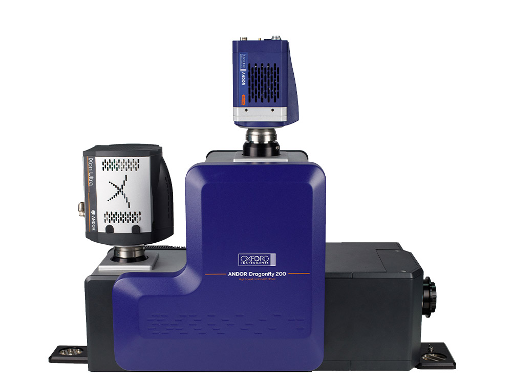
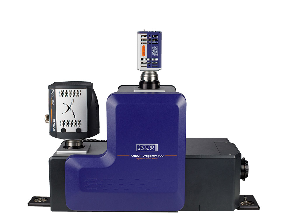
Discover our NEW Dragonfly 400 System
Deeper Imaging. Deeper Insights. Deeper Understanding.
Dragonfly spinning disk confocal delivers leading performance for industrial and academic users. Its modular design adds functionality and capability at each level.
Use the hotspots below to explore the key features and benefits of Dragonfly.

Dragonfly performs at a very high level and its ease of use allows researchers to acquire valuable data at speed. The system really excels for applications that are more demanding, such as high resolution live cell imaging.
Derek Toomre, PhD - Professor of Cell Biology at Yale School of Medicine.

Dragonfly is fast, sensitive, and fantastically flexible. We have 120 research groups working at our Core Facility every year, and they bring in everything you could imagine to image. Having the flexibility to switch between different modes and colors, and having a system that is very sensitive for single-molecule imaging through to spinning-disk confocal microscopy, is amazing for us.
Prof. Jeffrey Caplan - Director of Bioimaging and Associate Professor, University of Delaware.
Use microscope selector tool below to have an overview of the best Dragonfly for your application or technique
Filter by:
A flexible, multi-modal platform that offers confocal, laser based widefield, transmitted light and SRRF-Stream+ functionality in one system.
3D SMLM widefield imaging capabilities (DNA-PAINT). Spatial transcriptomics / large model organisms high-performance capabilities.
Fully configured Dragonfly system, adding 3D SMLM in widefield, confocal, B-TIRF (DNA-PAINT, dSTORM and PALM) and live cell TIRF imaging.
All Dragonfly imaging systems are compatible with Andor Photostimulation products. Please note that we indicate the best system for your application. In some cases, a lower model could deliver acceptable results. Please see our technical article selecting your Andor Microscope for more information.


© Oxford Instruments 2026
 Powered by Bioz
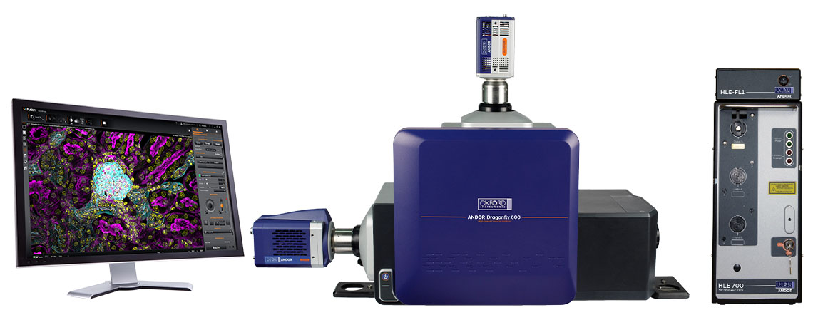
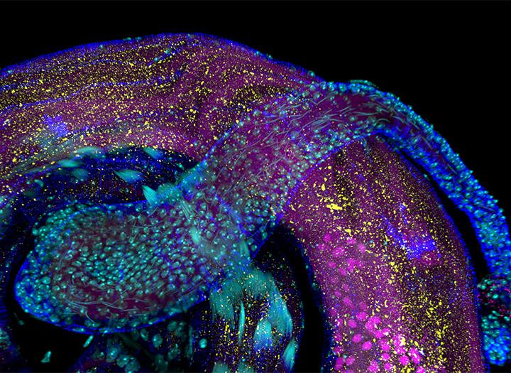
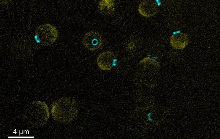
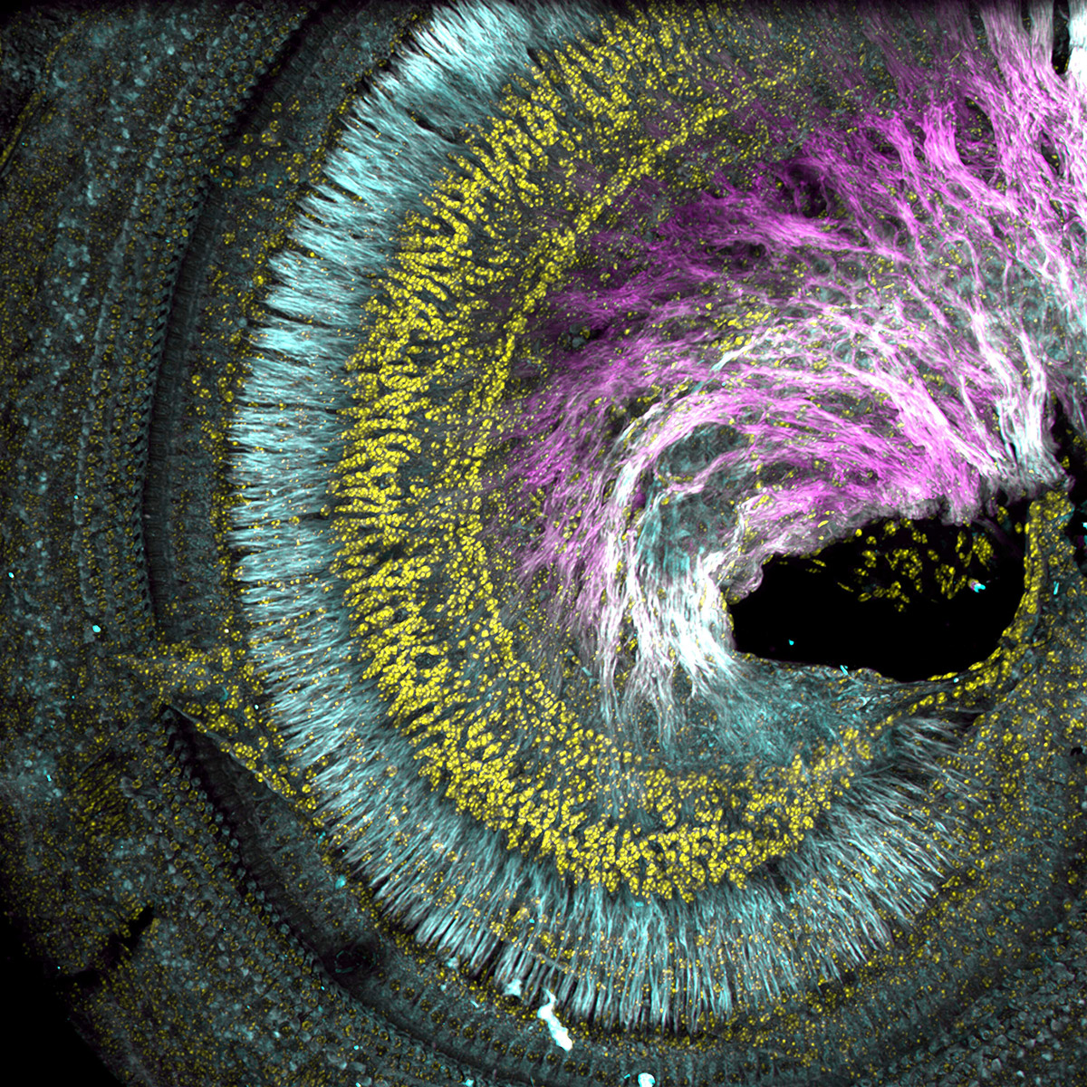
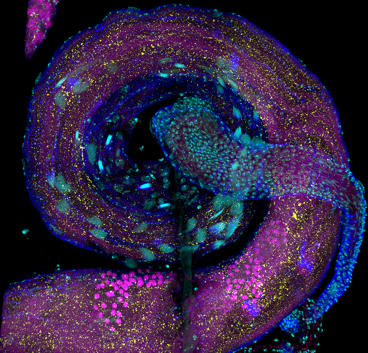
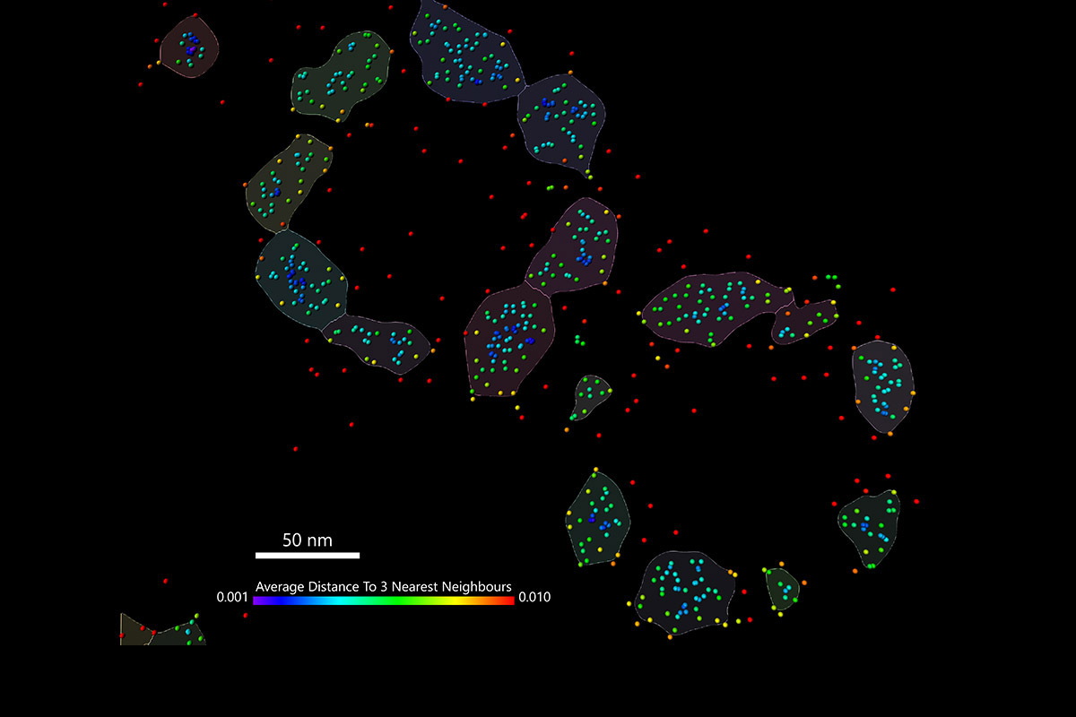
 Sample courtesy: Ching-Ho Chang, PhD; Harmit Malik Lab – Fred Hutchinson Cancer Research Center")