Resources
 Part of the Oxford Instruments Group
Expand
Collapse
Part of the Oxford Instruments Group
Application Notes
Author: Jinhui Guo, Benjamin Achatz, Matthias Zeilerbauer, Laerte L. Patera
Published: 01 May 2026 · Last updated: 26 May 2026
Porphyrins are highly conjugated macrocycles, capable of coordinating metal ions at the central cavity. Owing to their tunable electronic structure and rich photophysics, they are central to applications in catalysis, light harvesting, biomedicine, and molecular electronics¹. Among metalloporphyrins, 5,10,15,20-tetrakis(4-aminophenyl)porphyrin zinc(II) (ZnTAPP) is employed as an electron donor in artificial photosynthetic systems². This study focuses on the photoluminescence (PL) of ZnTAPP multilayer films thermally evaporated on an atomically clean Ag(111) surface, and compares the results with PL from solutions of ZnTAPP in dimethyl sulfoxide (DMSO). The objectives are to study emission peak positions, widths, and vibronic structure in solution and solid state, and to rationalize spectral shifts and lineshape changes in terms of temperature, solvation, and intermolecular interactions.
An Andor Kymera-193iA-SIL spectrograph optimized for the visible range was coupled to an Andor iDus DU420A-BEX2-DD back-illuminated CCD. The detector was operated at −80 °C to minimize dark current, thereby improving the signal-to-noise ratio for weak PL. An excitation wavelength of 450 nm (NKT SuperK FIANIUM 15, equipped with a SuperK VARIA variable tunable filter) was used for both the solution and multilayer film samples. For the PL measurements, we employed a 300 lines/mm grating (blaze 500 nm) and a 100 µm entrance slit, which provide a spectral resolution of about 2 nm (as calculated using the Andor resolution calculator: https://andor.oxinst.com/tools/resolution-calculator). The ZnTAPP multilayer film was measured at 77 K under ultra-high vacuum (UHV) for an integration time of 600s, whereas the ZnTAPP solution in DMSO was measured at 300 K under ambient conditions and an integration time of 10s.
Figure 1 shows the photoluminescence spectra of the ZnTAPP multilayer film and ZnTAPP in DMSO solution. Using a 450 nm excitation, both solution and film PL spectra exhibit a dominant emission peak centered near 650 nm, with a secondary feature around 690 nm, consistent with the vibronic progression of the Q-band emission from S1→S0. The PL spectra were normalized to the main peak to enable direct comparison of line shape and vibronic structure. Relative to the solution spectrum, the film spectrum exhibits a measurable blue shift, narrower linewidths, and more clearly resolved vibronic features. Instead, the solution spectrum at 300 K is substantially broader and more symmetric. In the film at 77 K, the vibronic peak at around 690 nm is reduced in intensity relative to the main peak compared to the solution spectrum, suggesting a more rigid environment of the ZnTAPP in the film compared to the solution. The broader, less-resolved PL in DMSO at RT can be attributed to increased homogeneous broadening. Thermal activation of nonradiative channels could also reduce the relative intensity of the 0–0 transition. Additionally, DMSO is a polar, high-permittivity solvent that differentially stabilizes the excited state relative to the ground state, increasing the Stokes shift and shifting emission to longer wavelengths via solvatochromism3. In contrast, the solid-state environment of the ZnTAPP multilayer at 77 K yields a smaller Stokes shift, sharper lines, and a blue-shifted emission.
Our measurements demonstrate that ZnTAPP shows pronounced environment- and temperature-dependent photoluminescence: transitioning from polar solution at 300 K to solid multilayers on Ag(111) at 77 K blue-shifts and sharpens the emission linewidth, and suppresses vibronic sidebands. Extending these measurements across temperature, film thickness, and substrate dielectric constant, together with time-resolved spectroscopy and absolute quantum-yield calibration, will enable quantitative extraction of Stokes shifts, Huang–Rhys factors, and excitonic coupling strengths, thereby informing rational design of porphyrin-based optoelectronic architectures.
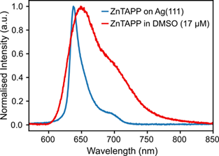
Photoluminescence spectra recorded under 450 nm excitation for (i) a 3 monolayer (3 ML) ZnTAPP film grown on Ag(111) measured at 77 K (blue trace) and (ii) a ZnTAPP solution in DMSO measured at 300 K (red trace). Both spectra were acquired with a 300 l/mm grating (blaze 500 nm).
[1] Osadchuk, I.; Aav, R.; Borovkov, V.; Clot, E. Chirogenesis in Zinc Porphyrins: Theoretical Evaluation of Electronic Transitions, Controlling Structural Factors and Axial Ligation. ChemPhysChem 2021, 22 (17), 1817–1833.
https://doi.org/10.1002/cphc.202100345.
[2] El-Khouly, M. E.; El-Mohsnawy, E.; Fukuzumi, S. Solar Energy Conversion: From Natural to Artificial Photosynthesis. J. Photochem. Photobiol. C Photochem. Rev. 2017, 31, 36–83.
https://doi.org/10.1016/j.jphotochemrev.2017.02.001.
[3] Lakowicz, J. R.; Masters, B. R. Principles of Fluorescence Spectroscopy, Third Edition. J. Biomed. Opt. 2008, 13 (2), 029901.
https://doi.org/10.1117/1.2904580

© Oxford Instruments 2026
