Resources
 Part of the Oxford Instruments Group
Expand
Collapse
Part of the Oxford Instruments Group

Buyers' Guides
Author: Judith Beer
Published: 03 Aug 2026 · Last updated: 06 Aug 2026
The illumination source plays a central role in fluorescence microscopy, as it determines compatibility with different imaging modes, spectral flexibility, image contrast, and resolution. In fluorescence microscopes, excitation stability and illumination uniformity are particularly important, as poor performance in these areas directly compromises image quality. This overview compares LED and laser options in benchtop fluorescence microscopes.
Benchtop fluorescence microscopes may integrate LEDs, lasers, or a combination of both. The choice of light source influences not only imaging performance, but also system cost and maintenance requirements.
* Xenon and metal halide lamps are not discussed further in this overview, as they are generally not utilised in modern systems and not relevant for modern benchtop imaging systems.
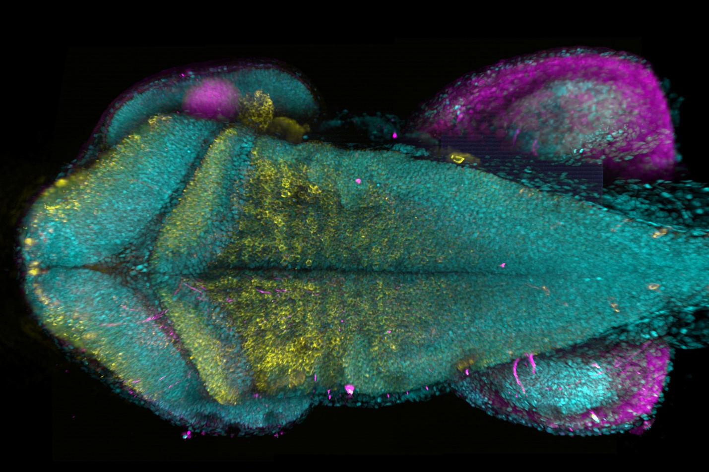
Fig. 1 – Image deep into the zebrafish brain with BC43. The combination of Oxford Instruments' patented Borealis illumination technology, dual microlens optics, and a highly accurate XZ stage enables acquisition of multiple XYZ planes throughout the zebrafish brain, which are seamlessly stitched into a single, continuous image. The image shows a maximum projection of zebrafish hindbrain at 48hpf staining the neurons (anti-HuC/D serum, yellow), glia (anti-zrf serum, magenta). 30 tiles were acquired to compose the image; each tile had 175 slices, over a 370 mm Z range. Image credits: Marco Campinho, Universidade do Algarve.
Learn more about the key technologies, components and practical considerations involved in selecting a benchtop fluorescence microscope.
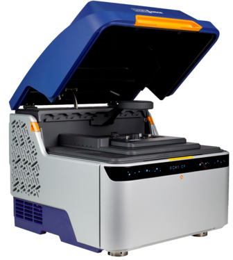
BC43 uses multimodal lasers integrated through the patented Borealis illumination system, ensuring uniform illumination across the field of view during imaging in the confocal mode.

© Oxford Instruments 2026