Why consider a Benchtop Fluorescence Microscope?
Benchtop fluorescence microscopes can offer advanced imaging capabilities without the size, cost, and complexity of a
traditional research microscope. High-performance systems such as the Oxford Instruments BC43 combine widefield
fluorescence imaging with options for confocal or even super-resolution. These features make benchtop platforms ideal
for cell biology, drug discovery, and education especially in space-limited labs or facilities with shared resources.
Who Benefits from a Benchtop System?
- Academic research labs performing routine imaging without reliance on shared full-scale systems
- Core facilities requiring an entry-level, easy-access system for users with standard imaging needs, not needing
specialised or high-end platforms
- Small labs or start-ups with limited space or budget but ongoing fluorescence imaging requirements
- Biotech & pharmaceutical labs supporting imaging for QC, assay validation, or sample screening
- Multi-user and teaching labs where ease of use and short training times are essential
How Much Do Capabilities Differ Between Benchtop Fluorescence Microscopes?
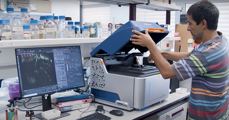
Benchtop fluorescence microscopes are not a single class of instruments but a broad and highly diverse category.
Their capabilities range from straightforward widefield setups for routine imaging to advanced systems incorporating
laser-based illumination, point scanning, spinning-disk confocal microscopy, and computationally enhanced
high‑resolution techniques. Because performance, speed, sensitivity, and feature sets vary considerably across this
spectrum, users must understand which specific capabilities align with their samples and experimental goals.
Key Questions to Ask Before Buying a Benchtop Microscope
- Which imaging modes do I need?
The required imaging modes depend on your sample type and research goals. Widefield fluorescence suits thin
specimens and routine imaging, whereas confocal capabilities enable optical sectioning for high-contrast and 3D
imaging of thicker samples. If your goal is to resolve structures beyond the diffraction limit, you require a system
offering super-resolution. Advanced transmitted light microscopy options help improve contrast in unstained samples.Read more
- What type of illumination source should I choose?
The illumination source defines a system’s suitability for advanced imaging modes and influences system cost and maintenance. LEDs are well-suited for most widefield workflows due to their stability, longevity, and gentle illumination. Confocal and super-resolution imaging require laser illumination for precise, high-intensity excitation. Ensure the supported wavelengths, if LEDs or lasers, match your current and future experimental needs. Read more
- Which objectives are best for my requirements?
Objectives must match your sample characteristics and imaging requirements. Magnification, numerical aperture (NA), and immersion type directly influence resolution, light collection, and imaging depth. High-NA objectives improve resolution but reduce working distance. For most accurate results, prioritise aberration-corrected objectives and high NA; for large sample imaging, be sure that the working distance matches your experimental and sample requirements.Read more
- What detector specifications should I look for?
Detector performance can vary significantly in signal sensitivity, image quality, and acquisition speed. Benchtop systems typically use integrated sCMOS cameras for image acquisition. Key parameters to compare camera performance are quantum efficiency (defines sensitivity), pixel size (impacts resolution and sensitivity), sensor size (determines field of view), dark current, and read noise (affect signal-to-noise ratio). Camera cooling further improves signal-to-noise ratios (see article: Is Cooling of an sCMOS Sensor Important).Read more
- Which other system features are important to accommodate my samples and experimental needs?
Stage design: If your workflow involves 3D imaging, large scans, multi-well plates, or time-lapse experiments, a motorised XYZ stage is essential. Read more
Sample handling: Confirm compatibility of your sample formats with the supported sample holders (including supporting software). Consider, slides, dishes, or multi-well plates.
Environmental control: For live-cell and long-term imaging studies, look for systems that support seamless integration of environmental enclosures or stage-top incubators to ensure optimal conditions and to maintain sample viability throughout the experiment. Read more
Vibrational control: Vibration control provides mechanical stability and is essential to ensure accurate imaging in time-lapse, 3D, or high-resolution imaging. Vibration control can be passive or active. Read more
- Advanced options – Is the system compatible with the requirements of high-containment laboratories (e.g., BSL-3 or BSL-4 facilities)?
Imaging in BSL-3 and BSL-4 laboratories is challenging due to strict containment, decontamination, and service requirements. Many researchers therefore image fixed samples outside the facility. For on-site imaging, it is important to verify that the microscope supports validated decontamination procedures and retains warranty and service coverage after decontamination.
Read more
Learn more about Benchtop Fluorescence microscopes
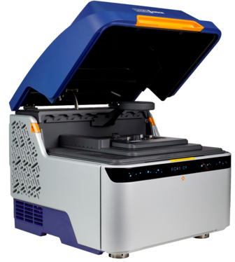
BC43 – Exceptional Performance, Certified Quality, High Productivity
- Versatile image modes in one system: Transmitted light, widefield, spinning-disc confocal, and SRRF-Stream+ super
resolution
- Modular design for in-field upgrade options
- Automated workflows with intuitive software
- 4 integrated laser lines for multi-colour fluorescence imaging
- High-sensitivity sCMOS camera
- Built-in vibration isolation and environmental control compatibility
- Certified quality control programs (IQ/OQ)
- Dedicated program for BSL-3 and BSL-4 laboratories
- Advanced image analysis with Imaris

 Part of the Oxford Instruments Group