Resources
 Part of the Oxford Instruments Group
Expand
Collapse
Part of the Oxford Instruments Group

Buyers' Guides
Author: Judith Beer
Published: 03 Aug 2026 · Last updated: 06 Aug 2026
Data acquisition and analysis software is as important as hardware functionalities in modern fluorescence microscopy. It determines how efficiently experiments can be configured, acquired, analysed and reproduced, with workflows ranging from routine imaging tasks to advanced multidimensional experiments.
The software capabilities offered by different benchtop fluorescence microscope systems can vary significantly. As a result, software often becomes a deciding factor when selecting a microscope, influencing not only workflow efficiency and ease of use but also the overall functionality and cost of the system.
This article reviews the key acquisition, analysis and automation capabilities to consider when comparing benchtop fluorescence microscopes, helping ensure that the system you choose can support your current and future imaging requirements.
Data acquisition software controls microscope hardware and defines how experiments are configured, automated, and recorded.
Many providers of benchtop systems offer advanced functionalities as optional, separately licensed modules. While this modularity allows high flexibility for complex experiments, it also means that total system costs can increase substantially once all required features are added. It is therefore important to carefully assess which software features match your application needs, which of them are included and which require additional licenses.
| Software Packages | Description |
| Basic packages |
|
| Advanced features |
|
| External device control |
|
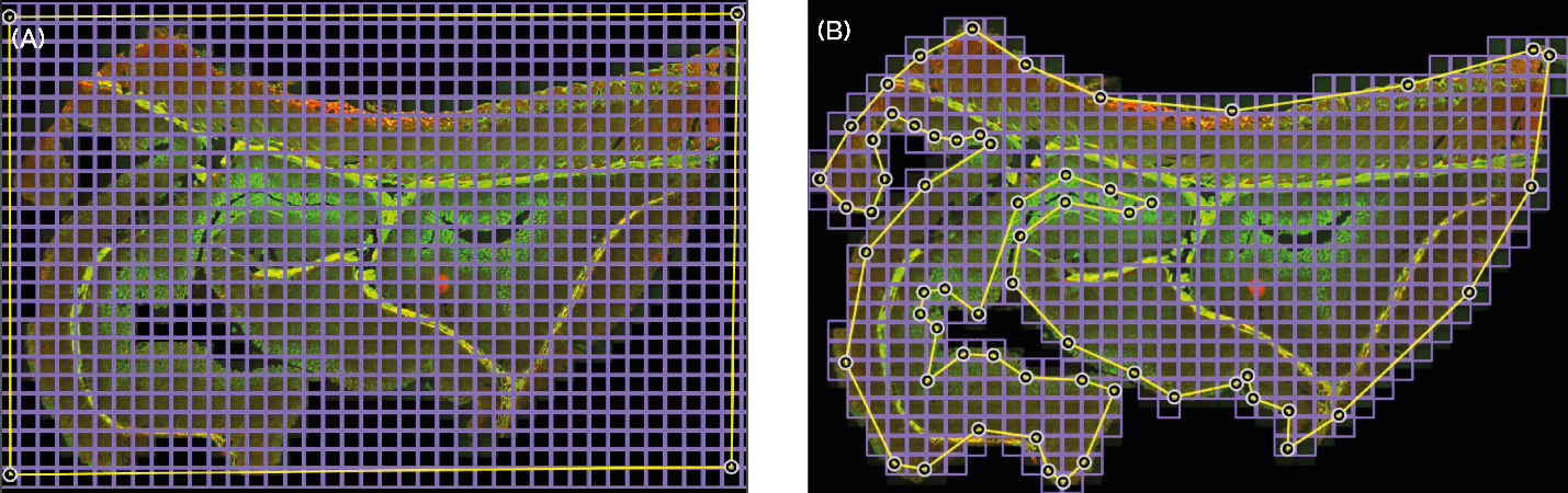
Fig. 1 — Irregular montage dramatically increases productivity in large sample imaging. The image compares a standard Montage (A) with an irregular montage acquired on the BC43 system (B). In the standard Montage, the acquisition area is defined by yellow boundaries, and the required tiles are shown in purple. Using this approach, the user must acquire 1040 tiles, resulting in 2080 images (2 channels). With the irregular montage applied to the same sample, only 646 tiles are required, corresponding to 1292 images. This represents a twofold increase in productivity for 2D imaging. The productivity gains become exponential when acquiring z stacks or additional imaging channels.
Efficient data processing is essential for final experimental results and conclusions once images are acquired. Data analysis typically includes data visualisation, segmentation, quantification, statistical evaluation, and interpretation.
As with data acquisition software, it is important to evaluate which analysis tools are provided with the microscope system. This includes understanding which features are included by default, which are offered as optional licensed modules, and how well they align with your applications, user expertise, and future needs.
Beyond system-included analysis tools, researchers may choose to process image data using external commercial software or open-source platforms.
| Software Type | Characteristics |
| Open-source solutions |
|
| Commercial platforms |
|
For a detailed comparison, see the article open-source vs paid image analysis software.
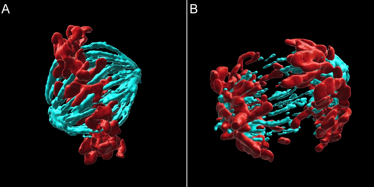
Fig. 2 — Mammalian cell division observed with the Oxford Instruments BC43 and rendered with Imaris. BC43 Widefield Imaging was used to image fixed mammalian cells. A) Metaphase cell, B) Anaphase cell. The images show 2 of 20 independent positions acquired in this experiment. Two channels were acquired per position across a range of 15 mm. Deconvolution was enabled in the protocol. Images were further processed in Imaris, presenting a maximum-intensity projection (MIP) of a surface-rendered image (Cyan – microtubules, Red – DNA). Image credits: Ines Baião-Santos, Álvaro Tavares – Universidade do Algarve; Claudia Florindo – Oxford Instruments
Automation features play an important role in improving ease of use, workflow efficiency, and measurement consistency in microscopy. The level of automation available varies widely across benchtop systems. Fully automated microscopes offer the highest degree of user-friendliness and standardisation, but they typically come with higher initial costs and, in some cases, reduced flexibility. Understanding which features are essential for your workflows helps balance usability, performance, flexibility, and cost.
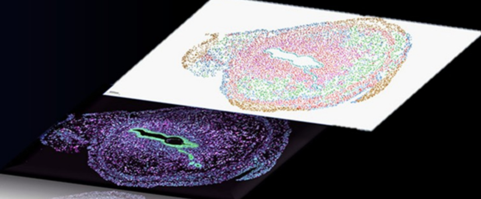
Fig. 3 — Spatial omics graphic representation. With the power of the REST API, users can orchestrate complex workflows by linking multiple protocols and external devices. Comprehensive gene-expression atlases can be generated by coordinating the spinning-disk confocal BC43 with automated microfluidic triggering.
Learn more about the key technologies, components and practical considerations involved in selecting a benchtop fluorescence microscope.

© Oxford Instruments 2026