Resources
 Part of the Oxford Instruments Group
Expand
Collapse
Part of the Oxford Instruments Group

Buyers' Guides
Author: Judith Beer
Published: 01 Jul 2026 · Last updated: 06 Aug 2026
The imaging mode describes the method a microscope uses to generate an image, defined by its illumination approach, detection strategy, and contrast mechanism. As each imaging mode differs in contrast, resolution, imaging depth, sensitivity, and speed, certain modes are better suited to specific sample types or experimental questions than others.
Benchtop fluorescence microscopes can vary widely in their capabilities and in the imaging modes they support. This overview explains the most common imaging modes available in benchtop systems and highlights the key factors to consider when evaluating a microscope.
Understanding which imaging modes are supported by a microscope is essential to ensure that the system can effectively address your biological or analytical goals. It is also important to note that imaging modes can vary significantly in technical complexity and cost, which can have a major impact on the overall price of the instrument.
Generates label-free contrast by collecting light passing through the sample. Commonly integrated into benchtop fluorescence microscopes for quick sample assessment, live-cell monitoring, focusing, and added structural context. Contrast performance and costs depend strongly on the specific technique.
| Transmitted-light technique | Description |
| Brightfield (BF) |
|
| Phase Contrast |
|
| Darkfield (DF) |
|
| Differential Interference Contrast (DIC) |
|
| Differential Phase Contrast (DPC) |
|
Illuminates the entire field of view simultaneously and collects both in-focus and out-of-focus emitted light. This is a fast, gentle, and cost-efficient imaging mode well-suited for routine fluorescence imaging and thin samples. However, it has limited depth resolution due to out-of-focus signal and does not suit thick and strongly scattering specimens.
Uses optical sectioning to improve contrast and resolution and allows for high-quality 3D-imaging. It requires laser-based illumination and pinholes to reject out-of-focus light.
| Confocal implementation | Description |
| Point-scanning confocal microscopy |
|
| Multi-point (spinning-disk) confocal microscopy |
|
Techniques that exceed the diffraction limit of conventional light microscopy (~200 nm, wavelength-dependent), enabling visualisation of structures down to ~10–150 nm, depending on the method. Hardware-based implementations are often complex and cost intensive. Not common in benchtop systems, but in some cases available.
| Super-resolution technique | Description |
| SIM (Structured Illumination Microscopy) |
|
| STED (Stimulated Emission Depletion Microscopy) |
|
| SMLM (Single-Molecule Localisation Microscopy techniques, e.g. STORM) |
|
| SRRF-Stream (Super-Resolution Radial Fluctuations) |
|
Multiple imaging modes available in a single benchtop platform enable flexibility across diverse samples and experimental workflows. This is typically associated with higher system costs. Modular systems may allow staged upgrades to scale functionality and investment over time.
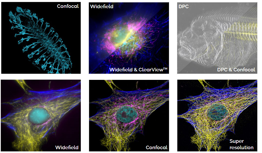
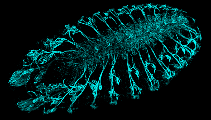
Fig. 1 — Representative images of imaging modalities available in BC43. Top images: (left) Confocal image of Drosophila embryos stained with a neuronal marker. (middle) Widefield & widefield-Clearview (deconvolution), mammalian cells showing actin, DNA, mitochondria, and microtubules. Images were deconvolved. (right) DPC and DPC image overlapped with a confocal image. Bottom images: The same cell was imaged using three different imaging modalities: widefield (left), confocal (centre), and super resolution (right). The increase in detail and resolution is clearly observed when different imaging modalities are used. Image credits: Alvaro Tavares and Marco Campinho, Universidade do Algarve, and Claudia Florindo, Oxford Instruments. Confocal (Drosophila) – CF & AT; DPC & Confocal – CF & MC; all others – CF.
Learn more about the key technologies, components and practical considerations involved in selecting a benchtop fluorescence microscope:
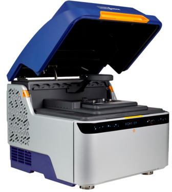
BC43 is a multimodal imaging system supporting transmitted light (brightfield and DPC), widefield, confocal imaging, and software-based super resolution (SRRF-Stream). The BC43 entry model featuring widefield imaging can be upgraded in the field to confocal with minimal system downtime, optional super-resolution can be added flexibly at any model (Widefield or confocal).

© Oxford Instruments 2026